By erasing distracting blood vessels from breast MRI scans, a new deep learning tool helps radiologists see hidden tumors more clearly.
Radiologists reading breast MRIs often face a crowded, confusing view. Bright, winding blood vessels frequently overlap with actual lesions, forcing doctors to guess where a tumor ends and healthy tissue begins. This visual clutter directly complicates surgical planning and biopsy targeting. When margins are unclear, clinicians may overestimate tumor size, leading to more invasive surgeries than necessary.
A new AI model called DeepVEST aims to solve this by digitally erasing these vascular distractions. This approach challenges the traditional reliance on raw projection images. It suggests that what we subtract from a medical scan is just as important as what we capture.
Clearing the visual noise
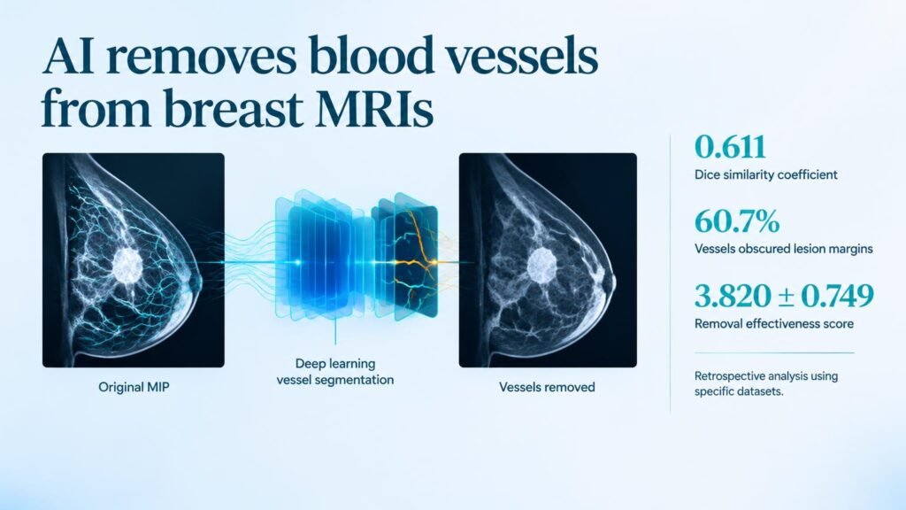
The scale of the problem is significant. In a reader study evaluating 150 assessments from the Duke and AMBL datasets, radiologists found that blood vessels partially or fully blocked lesion margins in 60.7% of cases. This is not a rare inconvenience. It is a daily clinical hurdle that slows down decision-making.
The model was developed using the Duke-Breast-Cancer-MRI dataset. By training on these complex cases, the algorithm learned to distinguish the rapid contrast uptake of blood vessels from the slower, more irregular pooling of contrast in malignant tumors.
DeepVEST tackled this by segmenting and removing the vessels. The AI achieved a Dice similarity coefficient of 0.611 for vessel segmentation. While that score reflects a moderate mathematical overlap, the practical utility in the clinic proved much higher. Five breast radiologists evaluated the results, confirming that the tool successfully cleared the field of view without destroying vital diagnostic data.
Key study metrics
- Vessels obscured lesion margins in 91 of 150 assessments.
- Radiologists rated vessel removal effectiveness at 3.820 out of 5.
- Artifacts appeared in 30.0% of cases, but with a very low severity score of 0.493 out of 5.
- The interreader agreement for removal quality was high, scoring a Gwet AC1 of 0.703.
The clinical reality check
This tool does not replace the radiologist. Instead, it alters their workflow by reducing cognitive fatigue. When the radiologists evaluated the system, they consistently agreed on its quality, yielding an artifact evaluation agreement of 0.736. This level of consensus is crucial for clinical adoption.
However, the technology has clear boundaries. A segmentation score of 0.611 means the AI still misses some vascular structures. Furthermore, the presence of artifacts in 45 of 150 scans means radiologists must remain vigilant. They cannot blindly trust the edited image, as they must ensure an AI-generated glitch is not mistaken for a real clinical feature.
Because this was a retrospective study, we still lack proof that this tool improves final patient outcomes or speeds up reading times. For now, the takeaway is practical. Subtractive AI is a viable path forward for medical imaging, proving that sometimes the best way to see a disease is to erase the healthy tissue surrounding it.
Read the full study in Radiology: Artificial Intelligence.