A new AI model proves that medical images contain hidden health data that vision-only tools completely miss.
Radiologists use CT scans to find immediate problems like fractures or tumors. But those same scans hold a quiet record of a patient’s aging, lab values, and future disease risks. A new model called Percival shows how vision-language models can extract this hidden layer of clinical reality without manual labeling.
This challenges the current trend of building narrow, task-specific AI tools. Instead of training one model to segment kidneys and another to find lung nodules, we can use a single model that understands the entire clinical context. It shifts the goal from simple image labeling to deep prognostic forecasting.
This shift has major operational consequences. Today, hospital IT departments must manage dozens of niche AI algorithms, which increases maintenance costs and integration friction. A single, generalizable model could streamline this pipeline by handling multiple clinical tasks at once.
How Percival works
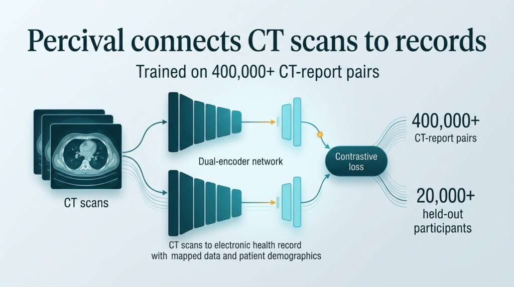
The model was trained on more than 400,000 CT-report pairs from the Penn Medicine BioBank. Researchers then tested the model on a held-out cohort of over 20,000 participants. This approach builds on previous efforts to link 3D imaging with text, such as the Merlin foundation model.
Percival uses a dual-encoder symmetric contrastive framework. This math matches the visual features of a CT scan to the descriptive text of the radiologist’s report. By doing this, the model’s internal map aligns naturally with demographic, physiological, and laboratory variation. It connects pixels to real-world patient outcomes.
Why text-guided AI wins
The researchers compared Percival against vision-only models and multi-organ segmentation tools. The results show that vision-language pretraining captures critical prognostic details that vision-only tools ignore. This finding aligns with other recent developments in the field, such as MG-3D’s knowledge-enhanced pre-training, which also highlights the value of text-guided learning.
- Analyzed data from over 20,000 held-out patients to map disease prevalence.
- Aligned imaging data with demographic, physiological, and laboratory variation.
- Outperformed vision-only contrastive models in longitudinal risk modeling.
Adding language to vision models gives them a clinical vocabulary they cannot learn from pixels alone. This allows the software to recognize patterns associated with chronic disease progression before they become visible to the naked eye.
The limits of prediction
The catch is that this study is retrospective and relies on a single health system’s biobank. We do not yet know how Percival performs when deployed in real-time clinical workflows or across diverse hospital networks with different scanner brands. Single-site success is not a guarantee of real-world reliability.
Health systems should not view these foundation models as immediate diagnostic replacements. Instead, they are powerful screening tools for population health. They can flag high-risk patients who need early intervention before symptoms appear.
Read the full study in npj Digital Medicine.