A new AI model can spot severe coronary blockage using a standard electrocardiogram, bypassing the need for expensive and radiation-heavy CT scans.
Why do we still inject dye and blast patients with radiation just to see if their arteries are clogged? Coronary CT angiography (CCTA) is the gold standard, but it requires heavy machinery, high costs, and patient stability. If a simple, cheap electrocardiogram (ECG) could map specific arterial blockages, the entire triage pipeline changes.
This study challenges the assumption that standard ECGs are too blunt to pinpoint vessel-specific damage. By predicting stenosis in four major coronary arteries, this AI foundation model shifts ECG from a general warning system to a precise anatomical map. It proves that deep learning can extract spatial, vessel-specific data from electrical signals that human cardiologists cannot see.
Mapping the blocked vessels
The researchers tested their model’s ability to predict severe stenosis in the four main coronary arteries. The model demonstrated strong diagnostic capabilities, though its accuracy varied significantly between the internal and external testing groups.
According to the study, the model achieved the following performance metrics:
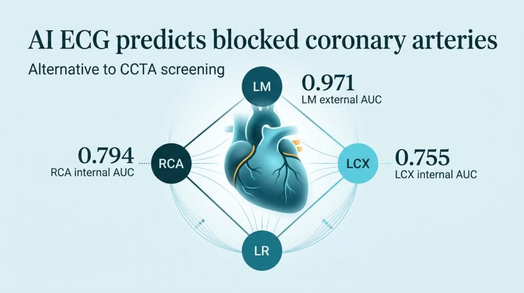
- Left main coronary artery (LM): Achieved an AUC of 0.818 internally and an outstanding 0.971 externally.
- Right coronary artery (RCA): Scored an AUC of 0.794 internally and 0.749 externally.
- Left circumflex artery (LCX): Reached an AUC of 0.755 internally and 0.727 externally.
- Left anterior descending artery (LAD): Logged an AUC of 0.744 internally and a lower 0.667 externally.
Hidden signals in normal scans
The real analytical breakthrough lies in the “normal” ECGs. The model maintained stable performance even in patients whose ECGs were flagged as clinically normal by doctors. This suggests the AI is reading sub-visual electrophysiological signatures of ischemia long before they manifest as classic abnormalities.
This builds on previous efforts to use deep learning for early detection. A 2023 study in Aging demonstrated the basic feasibility of ECG-based coronary screening. However, mapping specific vessels, rather than just general disease, represents a major leap forward. Another study in the European Heart Journal – Digital Health highlighted AI-ECG’s promise in stable angina, but this new foundation model takes the precision down to individual arterial branches.
The limits of prediction
We must look closely at the drop-off in the left anterior descending (LAD) artery performance. The external validation AUC fell to 0.667. The LAD is often called the “widowmaker” because blockages here are lethal. If the AI struggles to reliably flag this specific vessel in external clinics, clinicians cannot rely on it as a standalone diagnostic tool yet.
The external validation results also reveal a curious divergence. The model achieved a near-perfect AUC of 0.971 for the left main artery externally, up from 0.818 internally. This inconsistency suggests the model’s clinical utility may vary depending on the patient population and the specific artery diseased. AI is finding patterns, but we are still translating what those patterns mean biologically.
This tool matters because it changes the referral pipeline. Instead of sending every patient with chest pain for an expensive CCTA, clinics can use this AI to stratify risk immediately. It turns a ubiquitous, low-cost test into a highly specific anatomical screening tool.
This analysis is based on research published in npj Digital Medicine.