A new AI model uses advanced imaging to train simple electrocardiograms, proving that cheap tests can carry the predictive power of expensive scans.
Why do we still rely on expensive, hard-to-access cardiac MRIs when a simple ECG is sitting in every clinic? Cardiac MRIs provide unmatched detail of the heart, but they are expensive and rare. Most clinics rely on the humble, century-old electrocardiogram instead. For years, developers tried to make ECG models smarter on their own, but they hit a performance ceiling.
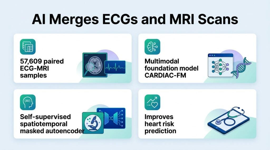
This new model, called CARDIAC-FM, challenges the assumption that cheap diagnostics must remain less accurate than advanced imaging. By training on both modalities, the AI learns to see the hidden structural signatures of an MRI within a standard, low-cost ECG. This shifts the focus of clinical AI from simple pattern recognition to cross-modal translation. It means we can distill the wisdom of rare, expensive scans into tools that any local clinic can use.
How the model was built
Researchers built CARDIAC-FM using 57,609 paired ECG and cardiac MRI samples. This massive dataset came from the UK Biobank, which has become a cornerstone for training large-scale cardiovascular models. The system uses a self-supervised masked autoencoder to align the electrical signals of the ECG with the spatial movement captured in the MRI scans. This allows the model to learn deep, shared representations of heart health.
The AI did not just memorize its training data. It generalized zero-shot to two completely different external cohorts: the Cardiovascular Health Study and the Multi-Ethnic Study of Atherosclerosis. Key performance results include:
- Improved prediction of incident atrial fibrillation and heart failure compared to existing ECG-only AI models.
- Successful risk prediction for additional outcomes including myocardial infarction, ischaemic stroke, and all-cause mortality.
- Strong performance even when deployed using ECG data alone, without requiring an active MRI scan.
Why this finding matters
This matters because it solves a major bottleneck in clinical AI deployment. Usually, multimodal models require all data types to be present to make a prediction, which is impractical in emergency rooms or rural clinics. CARDIAC-FM breaks this constraint. It allows a clinic with a basic ECG machine to benefit from the diagnostic insights of a high-end MRI scanner located hundreds of miles away.
However, the study has limitations. It relies on retrospective cohorts, and we do not yet know how the model handles the messy, real-world noise of everyday clinical ECGs. Furthermore, while the ECG-only mode is strong, the highest predictive gains still require actual cardiac MRI data. The digital divide in imaging access remains a factor, even if this software helps bridge the gap.
Ultimately, this approach shows that the future of medical AI is not about building bigger single-test models. It is about using rich, expensive data to elevate the tools we already have.
Read the full preprint on medRxiv.