
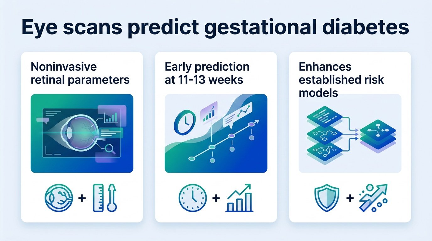
Adding retinal imaging to standard blood tests improves early gestational diabetes screening, but the incremental gain is smaller than AI optimists might hope.
Can we spot a metabolic crisis in the back of a pregnant patient’s eye months before it happens? Doctors usually wait until the 24th week of pregnancy to screen for gestational diabetes mellitus (GDM) using a time-consuming glucose tolerance test. By then, the metabolic disruption is already underway, leaving clinicians playing catch-up.
A new study attempts to bypass this delay by using machine learning to analyze the retinal blood vessels of pregnant women in their first trimester. The results complicate the popular narrative that AI-driven imaging can easily replace traditional blood work. Instead, the data shows that retinal features are not a magic shortcut, but rather a modest tuning knob. They refine risk stratification rather than rewriting the diagnostic playbook.
The predictive power of eyes
Researchers tracked 1,774 pregnant women, of whom 324 (or 18.3%) developed GDM. They built the LIGHT model using CatBoost, a machine learning algorithm, combining maternal factors, blood markers, and eight retinal features. The model identified the six most critical risk predictors: body mass index, triglyceride glucose index, age, HbA1c, and two eye metrics—standard deviation of angle deviation and angle-based tortuosity.
The performance numbers reveal a stark reality about relying on eye scans alone. The key model comparisons highlight this hierarchy:
- The LIGHT Model (Full): Achieved an AUC of 0.762, showing the strongest predictive power.
- The Glycolipid Model (No Eye Scans): Reached an AUC of 0.716 using only blood and maternal data.
- The Baseline Model: Managed an AUC of 0.687 using basic demographic data.
- The Eye Model (No Blood Tests): Performed poorly on its own, yielding an AUC of just 0.619.
This hierarchy shows that eye scans cannot replace blood tests. When compared directly to the Glycolipid model, the LIGHT model’s overall AUC improvement was not statistically significant. The eye is a window to the vascular system, but it is not a replacement for metabolic chemistry.
Why the nuance matters
This statistical flatline is where the analysis gets interesting. Why should clinics invest in retinal cameras if the overall diagnostic accuracy bump is negligible? The answer lies in how we classify patients who sit on the edge of a diagnosis.
The LIGHT model achieved a positive event net reclassification improvement (event-NRI) of 0.719. This means the eye scans successfully caught high-risk women whom standard blood tests would have missed. However, this came alongside a modest overall continuous NRI of -0.198 and an integrated discrimination improvement of -0.039.
For clinics, this is a trade-off. Retinal imaging is noninvasive and fast, but it requires specialized cameras and training. If the goal is to catch every possible case early, the extra hardware might be justified to reclassify those borderline patients. But we must be honest about the friction of adding eye scans to routine prenatal care when the diagnostic payoff remains incremental.
Read the full study in Cardiovascular Diabetology.