By predicting brain pressure from routine heart and blood signals, a new deep learning model challenges the necessity of invasive skull-drilling in intensive care units.
To measure the pressure inside a swollen brain, doctors must drill a hole through the skull to insert a physical catheter. It is a risky, invasive procedure reserved only for a narrow subset of the most critically ill patients. What if we could read the brain’s pressure waveform without ever touching the skull?
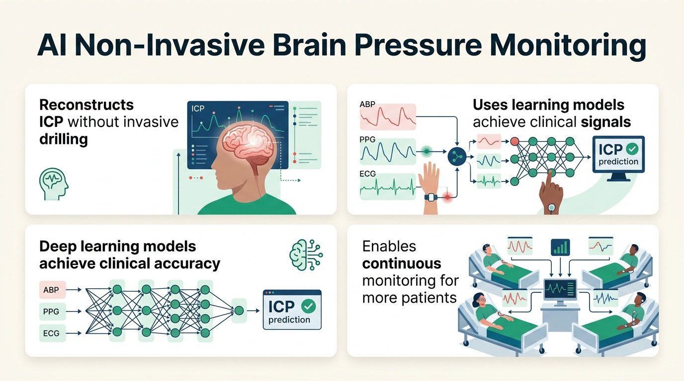
A new study suggests we can. By training AI on routine bedside signals like heart rate and blood pressure, researchers reconstructed intracranial pressure (ICP) waveforms with clinically relevant accuracy. This challenges a long-standing clinical assumption. We have acted as if the physical pressure inside the cranium is too isolated to be inferred from peripheral blood flow. This model proves that the systemic cardiovascular system carries a readable signature of brain pressure.
If validated, this shifts ICP monitoring from a high-risk surgical intervention to a software-enabled utility. It means clinicians could monitor brain swelling in patients who do not currently meet the strict, risky criteria for a skull bolt.
The data behind the model
The researchers developed and tested their models using a robust dataset from two major quaternary health systems. They analyzed data from 158 critically ill adults representing approximately 5,322 hours of continuous monitoring. The training and validation data came from Johns Hopkins Hospital in Baltimore and Beth Israel Deaconess Medical Center in Boston.
The team fed five different deep learning architectures three routine extracranial signals: high-frequency arterial blood pressure (ABP), photoplethysmography (PPG), and electrocardiography (ECG). They used actual invasive intraparenchymal ICP measurements as the ground truth. They evaluated two fusion strategies and three distinct training objectives to find the best fit.
What the AI achieved
The models did not just estimate a single pressure number. They reconstructed the entire, continuous pressure wave. The performance held up even when tested on an independent, external database. Here is how the different AI setups performed during validation:
- The gated recurrent model with late fusion achieved the lowest error, with a mean absolute error of 4.276 mmHg [95% CI 4.269, 4.283].
- The attention-based model with early fusion had the highest error of 4.946 mmHg [95% CI 4.938, 4.956].
- Waveform similarity, measured by Pearson correlation, ranged from 0.599 [95% CI 0.599, 0.600] to 0.722 [95% CI 0.722, 0.723].
- The multiscale encoder-decoder model demonstrated the most favorable balance between error and waveform correlation.
The real-world catch
An error margin of over 4 mmHg is not trivial in neurocritical care. In a patient hovering near the critical threshold of 20 mmHg, a 4 mmHg discrepancy could mean the difference between administering emergency therapies or doing nothing. Clinicians will need narrower error bounds before they trust software over a physical catheter.
Furthermore, the study relies on high-frequency arterial blood pressure lines. While less invasive than a skull bolt, an arterial line is still an invasive procedure. To truly democratize this technology, future models must prove they can work using only completely non-invasive inputs like standard finger-clip PPG and chest ECG leads. This work shows that the brain’s pressure signature is not locked behind the skull. It is written in the pulse.
Read the full preprint on medRxiv.