⚡ Quick Summary
This study explored a non-invasive radiomics-based machine learning algorithm to differentiate between upper urinary tract urothelial carcinoma (UTUC) and renal cell carcinoma (RCC) using preoperative computed tomography (CT) datasets. The model demonstrated a sensitivity of 88.4% and specificity of 81% in the training cohort, indicating its potential to enhance preoperative diagnostic workflows.
🔍 Key Details
- 📊 Dataset: 236 patients, with a median age of 70.5 years
- 🧩 Features used: Radiomic features extracted from CT imaging
- ⚙️ Technology: Machine learning model utilizing LASSO for feature selection
- 🏆 Performance: Training cohort: Sensitivity 88.4%, Specificity 81% (AUC: 0.93)
🔑 Key Takeaways
- 🔬 Radiomics can effectively differentiate between UTUC and RCC preoperatively.
- 💡 Machine learning enhances diagnostic accuracy compared to traditional imaging methods.
- 📈 Validation cohort showed a sensitivity of 80.6% and specificity of 80% (AUC: 0.87).
- 🔍 Subgroup analysis revealed robust performance in distinguishing clear cell RCC from high-grade UTUC.
- ⚠️ Limitations include the need for independent validation in future randomized controlled trials.
- 🌟 Potential to integrate this technology into current preoperative diagnostic workflows.
- 🗓️ Study conducted by a team of researchers from various institutions.
- 🆔 Clinical Trial Number: Local ethics committee no. 20-179.
📚 Background
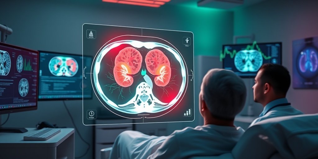
Differentiating between upper urinary tract urothelial carcinoma (UTUC) and renal cell carcinoma (RCC) is crucial for determining appropriate surgical interventions. Traditional imaging techniques often fall short in providing clear distinctions between these two types of cancers. The advent of radiomics—the extraction of large amounts of features from medical images—combined with machine learning offers a promising avenue for enhancing diagnostic accuracy in oncology.
🗒️ Study
This study retrospectively analyzed preoperative CT venous-phase datasets from patients diagnosed with either UTUC or RCC. The researchers manually segmented tumors and extracted radiomic features according to the International Image Biomarker Standardization Initiative. A predictive model was developed using the least absolute shrinkage and selection operator (LASSO), with the dataset divided into a training cohort (70%) and a test cohort (30%).
📈 Results
The model achieved impressive results, with a sensitivity of 88.4% and specificity of 81% in the training cohort, yielding an AUC of 0.93. In the validation cohort, the sensitivity was slightly lower at 80.6%, but specificity remained strong at 80% (AUC: 0.87). Notably, subgroup analyses indicated that the model performed well in distinguishing clear cell RCC from high-grade UTUC, with a sensitivity of 84% and specificity of 73.1%.
🌍 Impact and Implications
The findings from this study suggest that machine learning-based radiomics models can significantly improve the differentiation between UTUC and RCC in preoperative settings. By integrating such advanced technologies into the diagnostic workflow, healthcare professionals may enhance patient outcomes through more accurate preoperative assessments. This could lead to better-tailored surgical interventions and improved prognostic evaluations.
🔮 Conclusion
This research highlights the transformative potential of machine learning in oncology, particularly in the context of differentiating between complex cancer types using imaging data. As the field progresses, further validation through randomized controlled trials will be essential to solidify these findings and encourage widespread adoption in clinical practice. The future of cancer diagnostics looks promising with the integration of innovative technologies!
💬 Your comments
What are your thoughts on the use of machine learning in cancer diagnostics? We would love to hear your insights! 💬 Leave your comments below or connect with us on social media:
Radiomics-based differentiation of upper urinary tract urothelial and renal cell carcinoma in preoperative computed tomography datasets.
Abstract
BACKGROUND: To investigate a non-invasive radiomics-based machine learning algorithm to differentiate upper urinary tract urothelial carcinoma (UTUC) from renal cell carcinoma (RCC) prior to surgical intervention.
METHODS: Preoperative computed tomography venous-phase datasets from patients that underwent procedures for histopathologically confirmed UTUC or RCC were retrospectively analyzed. Tumor segmentation was performed manually, and radiomic features were extracted according to the International Image Biomarker Standardization Initiative. Features were normalized using z-scores, and a predictive model was developed using the least absolute shrinkage and selection operator (LASSO). The dataset was split into a training cohort (70%) and a test cohort (30%).
RESULTS: A total of 236 patients [30.5% female, median age 70.5 years (IQR: 59.5-77), median tumor size 5.8 cm (range: 4.1-8.2 cm)] were included. For differentiating UTUC from RCC, the model achieved a sensitivity of 88.4% and specificity of 81% (AUC: 0.93, radiomics score cutoff: 0.467) in the training cohort. In the validation cohort, the sensitivity was 80.6% and specificity 80% (AUC: 0.87, radiomics score cutoff: 0.601). Subgroup analysis of the validation cohort demonstrated robust performance, particularly in distinguishing clear cell RCC from high-grade UTUC (sensitivity: 84%, specificity: 73.1%, AUC: 0.84) and high-grade from low-grade UTUC (sensitivity: 57.7%, specificity: 88.9%, AUC: 0.68). Limitations include the need for independent validation in future randomized controlled trials (RCTs).
CONCLUSIONS: Machine learning-based radiomics models can reliably differentiate between RCC and UTUC in preoperative CT imaging. With a suggested performance benefit compared to conventional imaging, this technology might be added to the current preoperative diagnostic workflow.
CLINICAL TRIAL NUMBER: Local ethics committee no. 20-179.
Author: [‘Marcon J’, ‘Weinhold P’, ‘Rzany M’, ‘Fabritius MP’, ‘Winkelmann M’, ‘Buchner A’, ‘Eismann L’, ‘Jokisch JF’, ‘Casuscelli J’, ‘Schulz GB’, ‘Knösel T’, ‘Ingrisch M’, ‘Ricke J’, ‘Stief CG’, ‘Rodler S’, ‘Kazmierczak PM’]
Journal: BMC Med Imaging
Citation: Marcon J, et al. Radiomics-based differentiation of upper urinary tract urothelial and renal cell carcinoma in preoperative computed tomography datasets. Radiomics-based differentiation of upper urinary tract urothelial and renal cell carcinoma in preoperative computed tomography datasets. 2025; 25:196. doi: 10.1186/s12880-025-01727-9