⚡ Quick Summary
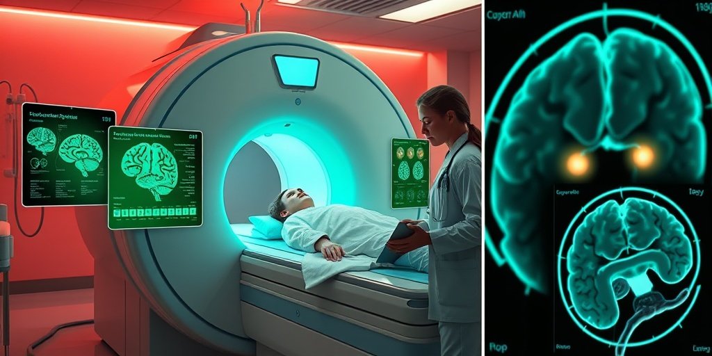
This study presents a novel approach for early brain tumor detection by utilizing Berkeley wavelet transformation (BWT) for segmenting MRI images. The method demonstrates improved accuracy and efficiency compared to existing techniques, highlighting its potential in clinical settings.
🔍 Key Details
- 🧠 Focus: Early detection of brain tumors
- 🖼️ Technology: MRI images and Berkeley wavelet transformation
- ⚙️ Classification Method: Support Vector Machine (SVM)
- 🔍 Morphological Operations: Erosion and dilation for edge detection
🔑 Key Takeaways
- 🧠 Brain tumors are a leading cause of mortality, making early detection crucial.
- 💻 Computer vision technologies can significantly enhance the detection process.
- 📊 The proposed method utilizes BWT for effective image segmentation.
- ⚙️ SVM is employed to classify tumor-affected and non-affected areas.
- 🔬 Morphological operations like erosion and dilation improve edge detection.
- 📈 Experimental results indicate superior accuracy and efficiency over existing methods.
- 🌍 Potential applications in clinical settings could lead to better patient outcomes.

📚 Background
Brain tumors pose a significant challenge in medical diagnostics, often leading to delayed treatment and poor patient outcomes. Traditional methods of tumor detection rely heavily on the expertise of clinical professionals, which can be both time-consuming and prone to human error. The integration of computer vision and advanced imaging techniques offers a promising solution to enhance the accuracy and speed of brain tumor detection.
🗒️ Study
The study conducted by Santhakumar et al. focuses on developing a method that leverages Berkeley wavelet transformation to segment MRI images for brain tumor detection. The researchers aimed to create a more efficient and accurate system that could assist clinicians in identifying tumors that cross the blood-brain barrier (BBB). The methodology involved converting images to greyscale, applying morphological operations, and utilizing SVM for classification.
📈 Results
The results of the study demonstrated that the proposed method significantly outperformed existing approaches in terms of accuracy and efficiency. The use of BWT for segmentation, combined with SVM classification, yielded promising results, indicating a high level of precision in distinguishing between tumor-affected and non-affected areas. Graphical representations of the experimental results further illustrated the effectiveness of the method.
🌍 Impact and Implications
The implications of this study are profound, as it suggests that integrating advanced imaging techniques with computer vision can revolutionize the way brain tumors are detected. By improving early detection rates, this method could lead to timely interventions and better patient outcomes. The potential for broader applications in clinical settings is immense, paving the way for enhanced diagnostic tools in neurology.
🔮 Conclusion
This research highlights the transformative potential of technology in the field of brain tumor detection. By employing Berkeley wavelet transformation and SVM classification, healthcare professionals can achieve more accurate and efficient diagnoses. As we continue to explore the integration of AI and imaging technologies in medicine, the future looks promising for improving patient care and outcomes in neurology.
💬 Your comments
What are your thoughts on this innovative approach to brain tumor detection? We would love to hear your insights! 💬 Leave your comments below or connect with us on social media:
Enhanced Early Brain Tumor Detection Crossing Blood-Brain Barrier through MRI Images Using Berkeley Wavelet-Transformation-Based Segmentation.
Abstract
Brain tumor is one of the reasons for several mortality cases in hospitals. Early detection and diagnosis of brain tumors are necessary to cure the disease early. The extraction of the tumor from the brain’s magnetic resonance image (MRI) is considered to be a difficult task when done by clinical experts, and it is also pretty time-consuming. These drawbacks can be overcome by using computer vision-based technologies. The proposed method detects brain tumor crossing the blood-brain barrier (BBB) through MRI images by using Berkeley wavelet transformation (BWT) for segmenting the affected areas. Support vector machine (SVM) is used for classification purpose by which the classification process is divided into two different categories namely, the tumor affected and tumor non-affected parts. Initially, the acquired image is converted to a greyscale from RGB. Then, image segmentation is done. During the image segmentation, morphological operations are carried out. Two morphological operations have been used in the proposed system. They are erosion and dilation. Both these techniques are used for edge detection. In erosion, the pixels are removed from the edges of the tumor image. In dilation, pixels are added at the edges of the tumor images. After the morphological operation, feature extraction is carried out. The features like homogeneity, contrast of the image and the energy might be determined. Then, the image is classified using the SVM classification algorithm. The experimental results have been tabulated and depicted using graphical representations. Comparing to the existing approaches the proposed method is proved to be better in accuracy and efficiency.
Author: [‘Santhakumar D’, ‘Prasanna R’, ‘Sivakumar M’, ‘Aswath S’, ‘Arthy PS’, ‘Kanna RR’]
Journal: Crit Rev Ther Drug Carrier Syst
Citation: Santhakumar D, et al. Enhanced Early Brain Tumor Detection Crossing Blood-Brain Barrier through MRI Images Using Berkeley Wavelet-Transformation-Based Segmentation. Enhanced Early Brain Tumor Detection Crossing Blood-Brain Barrier through MRI Images Using Berkeley Wavelet-Transformation-Based Segmentation. 2025; 42:93-125. doi: 10.1615/CritRevTherDrugCarrierSyst.2025055526