⚡ Quick Summary
This study developed a deep learning model to automatically segment the extrapancreatic nerve plexus and diagnose extrapancreatic perineural invasion (EPNI) in patients with pancreatic ductal adenocarcinoma (PDAC). The model demonstrated an accuracy of 79.7% in the training set and 72% in the validation set, highlighting its potential as a diagnostic tool.
🔍 Key Details
- 📊 Dataset: 332 patients diagnosed with PDAC
- 🧩 Features used: Enhanced CT scans
- ⚙️ Technology: nnUNet network with attention mechanism
- 🏆 Performance: Accuracy: 79.7% (training), 72% (validation)
🔑 Key Takeaways
- 📊 EPNI is a significant prognostic factor in PDAC, often leading to positive resection margins.
- 🤖 Deep learning can enhance the segmentation and diagnosis of EPNI.
- 🧠 The model achieved a Dice similarity coefficient of 60% for the celiac axis nerve plexus.
- 📈 The model’s ROC curve areas were 0.8 in training and 0.85 in validation, indicating strong diagnostic capability.
- 🔍 Further studies are needed to refine the model’s performance.
- 🌟 This research represents a promising step towards automated diagnostic tools in oncology.
📚 Background
Pancreatic ductal adenocarcinoma (PDAC) is a highly aggressive cancer with a poor prognosis. One of the challenges in managing PDAC is the presence of extrapancreatic perineural invasion (EPNI), which complicates surgical resection and is associated with worse outcomes. Traditional diagnostic methods can be invasive and subjective, necessitating the development of more reliable, automated techniques.
🗒️ Study
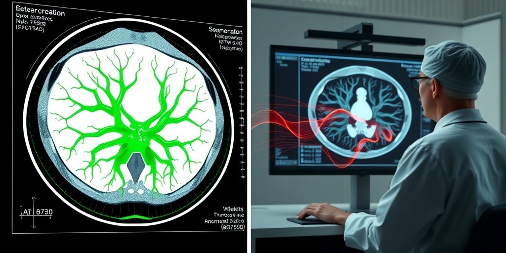
Conducted between August 2018 and December 2022, this retrospective study enrolled patients diagnosed with PDAC who underwent enhanced CT scans. The researchers aimed to create a fully automatic model for segmenting the extrapancreatic nerve plexus and diagnosing EPNI using advanced deep learning techniques, specifically the nnUNet network combined with an attention mechanism.
📈 Results
The study included a total of 332 patients, with 53.3% diagnosed with EPNI. The model demonstrated modest performance in segmenting the nerve plexus, achieving Dice similarity coefficients of 60% for the celiac axis, 68.2% for the superior mesenteric artery, and 35.7% for the common hepatic artery. However, it excelled in diagnosing EPNI, with an accuracy of 79.7% in the training set and 72% in the validation set, alongside ROC curve areas of 0.8 and 0.85, respectively.
🌍 Impact and Implications
The findings from this study could significantly impact the field of oncology by providing a novel and automated diagnostic tool for EPNI in PDAC patients. By improving the accuracy of diagnosis, this model has the potential to enhance surgical planning and patient outcomes. As the healthcare industry increasingly embraces artificial intelligence, such advancements could lead to more personalized and effective treatment strategies.
🔮 Conclusion
This study highlights the transformative potential of deep learning in the diagnosis of EPNI in pancreatic cancer. While the model shows promise, further research is essential to refine its capabilities and validate its clinical utility. The integration of AI in diagnostic processes could pave the way for improved patient care and outcomes in oncology.
💬 Your comments
What are your thoughts on the use of deep learning in cancer diagnostics? We would love to hear your insights! 💬 Leave your comments below or connect with us on social media:
Deep learning to predict extrapancreatic perineural invasion at CT images.
Abstract
BACKGROUND: Extrapancreatic perineural invasion (EPNI) was an adverse prognostic factor in patients with pancreatic ductal adenocarcinoma (PDAC) and responsible for positive resection margin. This study aimed to develop an automatic model for segmenting the extrapancreatic nerve plexus and diagnosing EPNI.
METHODS: In this retrospective study, patients diagnosed with PDAC who underwent enhanced computer tomography scans between August 2018 and December 2022 were enrolled. These cases were divided into training sets with radiological EPNI labels, and validation sets with pathological EPNI labels. The extrapancreatic nerve plexus was segmented first via the nnUNet network and attention mechanism under the background of segmentation of PDAC and adjacent vessels. A 2D classifier was applied to diagnose EPNI based on the segmentation of the nerve plexus. The Dice similarity coefficients (DSCs), receiver operating characteristic (ROC) curve, and diagnostic accuracy were employed to evaluate the performance of the model.
RESULTS: A total of 332 consecutive patients were enrolled and classified into the training (n = 282) and validation (n = 50) sets. Patients diagnosed with EPNI accounted for 177 of the 332 patients (53.3%). On the one hand, the model showed modest DSCs in segmenting nerve plexus around celiac axis (CA), superior mesenteric artery (SMA), and common hepatic artery (CHA), which were 60, 68.2 and 35.7%, respectively. On the other hand, the model had a favorable performance in diagnosing EPNI; the accuracy and areas under the ROC curve were 0.797, 0.8 in training set and 0.72, 0.85 in the validation set.
CONCLUSIONS: The fully automatic deep learning model for segmenting the nerve plexus and diagnosing EPNI was a novel and promising tool. Further studies are required to improve the model performance.
Author: [‘Cai Z’, ‘Zou L’, ‘Li Q’, ‘Chen J’, ‘Qiu Y’, ‘Ji J’, ‘Mao L’]
Journal: Ann Med
Citation: Cai Z, et al. Deep learning to predict extrapancreatic perineural invasion at CT images. Deep learning to predict extrapancreatic perineural invasion at CT images. 2025; 57:2568116. doi: 10.1080/07853890.2025.2568116