⚡ Quick Summary
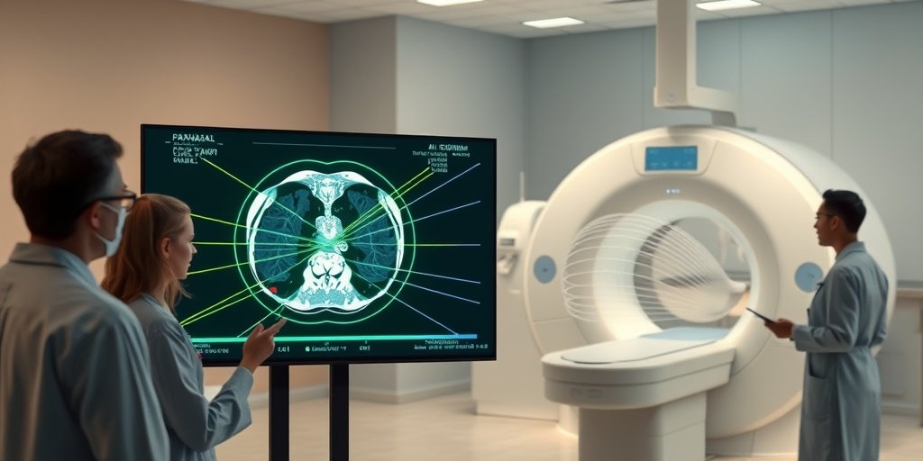
A recent study developed a deep learning approach for the rapid diagnosis of mucormycosis using paranasal CT images. The ConvNeXt Small model achieved an impressive 100% accuracy, demonstrating the potential of AI in expediting the diagnostic process for this life-threatening fungal infection.
🔍 Key Details
- 📊 Dataset: 794 CT images from patients with mucormycosis, nasal polyps, or normal findings
- ⚙️ Technology: Deep learning models (ResNet50 and ConvNeXt Small)
- 🏆 Performance: ConvNeXt Small: 100% accuracy; ResNet50: 99.16% accuracy
- 🔄 Methodology: 70/30 train-test split with five-fold cross-validation
🔑 Key Takeaways
- 🧠 Deep learning can significantly enhance the speed and accuracy of mucormycosis diagnosis.
- 💡 ConvNeXt Small model achieved a remarkable 100% accuracy in detecting mucormycosis.
- 📈 ResNet50 also performed well with 99.16% accuracy.
- 🔍 Cross-validation confirmed consistent results, indicating no overfitting.
- 🧪 Transfer learning proved beneficial, as training models from scratch resulted in lower accuracy.
- ⚠️ Models are adjuncts to traditional biopsy methods, not replacements.
- 🌐 Future research should focus on validating these models on external datasets.
- ⏳ Early intervention in mucormycosis could be facilitated through these AI tools.
📚 Background
Mucormycosis is a serious fungal infection that poses a significant threat to patients, particularly those with compromised immune systems. Traditional diagnostic methods often rely on invasive procedures like biopsies, which can delay treatment. The integration of artificial intelligence into diagnostic processes offers a promising avenue for rapid and non-invasive detection, potentially saving lives.
🗒️ Study
This retrospective study analyzed 794 CT images from patients diagnosed with mucormycosis, nasal polyps, or normal findings. The researchers employed two advanced deep learning models, ResNet50 and ConvNeXt Small, to classify the images into three categories. The study utilized a 70/30 train-test split and implemented five-fold cross-validation to ensure robust performance evaluation.
📈 Results
The results were striking: the ConvNeXt Small model achieved a flawless 100% accuracy on the test set, with precision, recall, and F1-score all at 1.00 for all classes. In contrast, the ResNet50 model achieved a commendable 99.16% accuracy, with precision and recall values around 0.99. The consistency of results across cross-validation further validated the models’ reliability.
🌍 Impact and Implications
The implications of this study are profound. By utilizing deep learning models for the diagnosis of mucormycosis, healthcare providers can potentially flag suspected cases for prompt treatment, thereby improving patient outcomes. These models could serve as rapid screening tools that complement traditional diagnostic methods, paving the way for earlier interventions and better management of this life-threatening condition.
🔮 Conclusion
This study highlights the transformative potential of AI in the field of medical diagnostics, particularly for conditions like mucormycosis. The ability to accurately and non-invasively detect such infections using CT scans could revolutionize the diagnostic landscape. As we look to the future, further validation and integration of these models into clinical workflows will be essential for enhancing patient care.
💬 Your comments
What are your thoughts on the use of AI for diagnosing mucormycosis? We’d love to hear your insights! 💬 Share your comments below or connect with us on social media:
Automated Mucormycosis Diagnosis from Paranasal CT Using ResNet50 and ConvNeXt Small.
Abstract
PURPOSE: Mucormycosis is a life-threatening fungal infection, where rapid diagnosis is critical. We developed a deep learning approach using paranasal computed tomography (CT) images to test whether mucormycosis can be detected automatically, potentially aiding or expediting the diagnostic process that traditionally relies on biopsy.
METHODS: In this retrospective study, 794 CT images (from patients with mucormycosis, nasal polyps, or normal findings) were analyzed. Images were resized and augmented for training. Two transfer learning models (ResNet50 and ConvNeXt Small) were fine-tuned to classify images into the three categories. We employed a 70/30 train-test split (with five-fold cross-validation) and evaluated performance using accuracy, precision, recall, F1-score, and confusion matrices.
RESULTS: The ConvNeXt Small model achieved 100% accuracy on the test set (precision/recall/F1-score = 1.00 for all classes), while ResNet50 achieved 99.16% accuracy (precision ≈0.99, recall ≈0.99). Cross-validation yielded consistent results (ConvNeXt accuracy ~99% across folds), indicating no overfitting. An ablation study confirmed the benefit of transfer learning, as training ConvNeXt from scratch led to lower accuracy (~85%) Conclusions: Our findings demonstrate that deep learning models can accurately and non-invasively detect mucormycosis from CT scans, potentially flagging suspected cases for prompt treatment. These models could serve as rapid screening tools to complement standard diagnostic methods (histopathology), although we emphasize that they are adjuncts and not replacements for biopsy. Future work should validate these models on external datasets and investigate their integration into clinical workflows for earlier intervention in mucormycosis.
Author: [‘Toprak SF’, ‘Dedeoğlu S’, ‘Kozan G’, ‘Ayral M’, ‘Can Ş’, ‘Türk Ö’, ‘Akdağ M’]
Journal: Bioengineering (Basel)
Citation: Toprak SF, et al. Automated Mucormycosis Diagnosis from Paranasal CT Using ResNet50 and ConvNeXt Small. Automated Mucormycosis Diagnosis from Paranasal CT Using ResNet50 and ConvNeXt Small. 2025; 12:(unknown pages). doi: 10.3390/bioengineering12080854