Overview
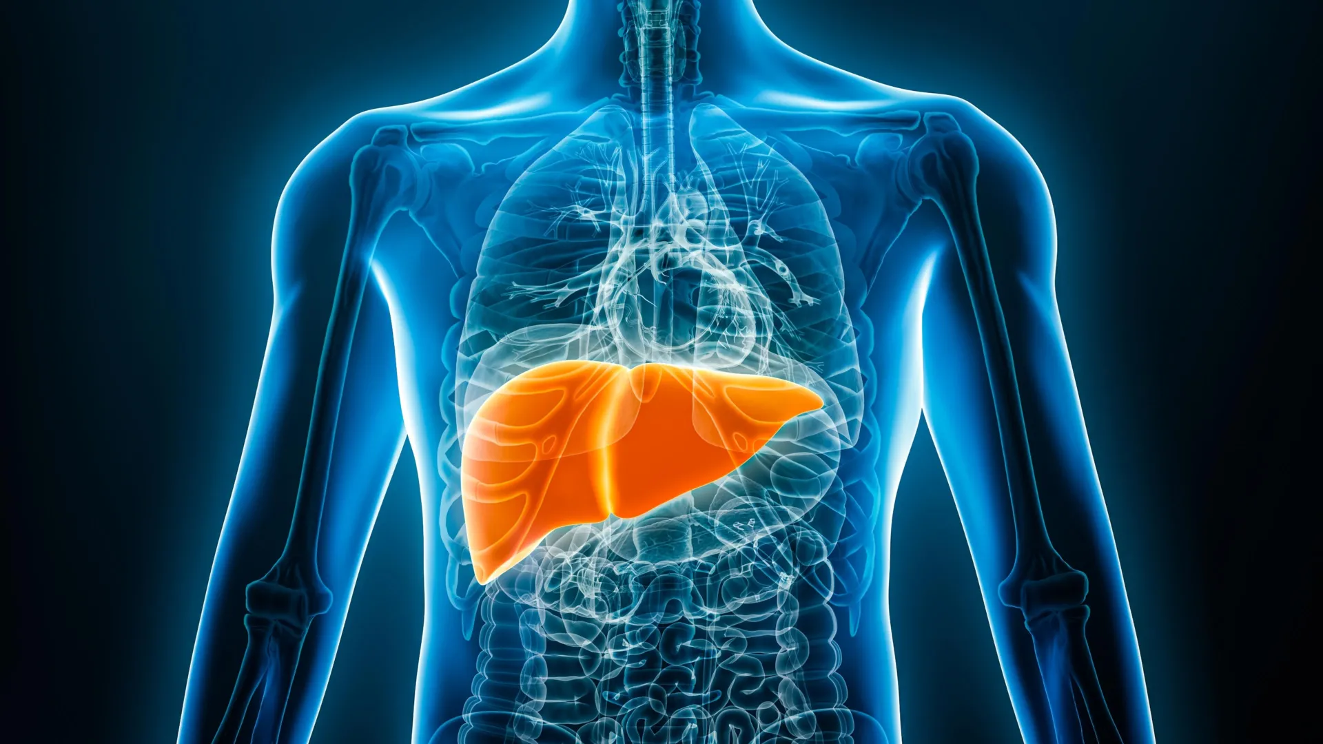
Fatty liver disease, characterized by fat accumulation in the liver, affects approximately one in four individuals globally. Early detection is vital to prevent severe complications such as cirrhosis and liver cancer.
Current Diagnostic Methods
Traditional diagnostic methods for fatty liver disease include:
- Ultrasounds
- CT scans
- MRIs
These methods require expensive equipment and specialized facilities. In contrast, chest X-rays are more commonly performed, cost-effective, and involve lower radiation exposure. Although primarily used for assessing lung and heart conditions, chest X-rays can also reveal signs of fatty liver disease.
Research Development
A research team from Osaka Metropolitan University, led by Associate Professors Sawako Uchida-Kobayashi and Daiju Ueda, has developed an AI model capable of detecting fatty liver disease from chest X-ray images. This study utilized:
- 6,599 chest X-ray images
- Data from 4,414 patients
The AI model was trained using controlled attenuation parameter (CAP) scores and demonstrated high accuracy, with an area under the receiver operating characteristic curve (AUC) between 0.82 and 0.83.
Future Implications
Professor Uchida-Kobayashi expressed optimism about the potential of this diagnostic method, stating, “The development of diagnostic methods using easily obtainable and inexpensive chest X-rays has the potential to improve fatty liver detection. We hope it can be put into practical use in the future.”
Reference
Ueda D, Uchida-Kobayashi S, Yamamoto A, Walston SL, Motoyama H, Fujii H, Watanabe T, Miki Y, Kawada N. Performance of a Chest Radiograph-based Deep Learning Model for Detecting Hepatic Steatosis. Radiol Cardiothorac Imaging. 2025 Jun;7(3):e240402. doi: 10.1148/ryct.240402