
Automated tissue analysis on CTA scans predicts post-procedure mortality in minutes, addressing a major bottleneck in cardiac risk assessment.
Manual quantification of aortic valve tissue before transcatheter aortic valve replacement is labor-intensive and variable. Echocardiography, the traditional standard, produces discordant severity ratings in roughly one-third of cases.
Speed Meets Prognostics
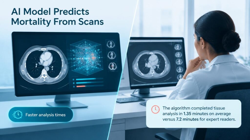
A deep-learning algorithm processed CT angiography tissue characterization in 1.35 minutes on average, compared to 7.2 minutes for expert readers. Beyond speed, the model independently predicted long-term all-cause mortality following TAVR. It extracts calcific and fibrotic tissue volume without human fatigue.
What Changes
Faster imaging analysis does not automatically replace clinicians or guarantee better patient outcomes. However, replacing manual tracing with a reproducible biomarker standardizes pre-procedural risk assessment.
The immediate takeaway is operational. Hospitals can automate routine CTA quantification to reduce radiologist workload while improving prognostic consistency for high-risk cardiac interventions.