
A digital stethoscope powered by an ensemble neural network outperforms expert cardiologists at spotting hidden left ventricular dysfunction before an echocardiogram.
Cardiologists relying on standard bedside acoustic checks miss low ejection fraction far too often. In a 100-patient reader substudy, human experts correctly identified left ventricular dysfunction with just 62.0% accuracy during physical examinations. That diagnostic gap leaves asymptomatic heart failure undetected until structural damage worsens.
That failure rate challenges the clinical value of unassisted bedside physicals. This trial proves that algorithmic interpretation of acoustic and electrical signals creates a reliable filter for echocardiography, shifting how clinics triage high-risk cardiac patients.
Outperforming Expert Clinical Consensus
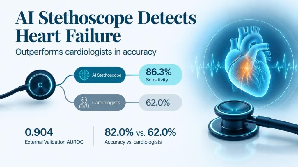
Researchers built an ensemble model combining a transformer network with a one-dimensional convolutional neural network. The algorithm analyzes four-site digital auscultation recordings paired with simultaneous single-lead ECG signals. In a 100-patient head-to-head trial, the AI achieved 82.0% accuracy compared to 62.0% for cardiologist consensus (P=0.004).
The system was trained on a multicenter cohort of 2,359 patients (mean age 69.7 years, 40.4% female, 8.9% with ejection fraction 40% or lower). It easily beat ECG-only tools and standard clinical logistic models incorporating B-type natriuretic peptide levels.
The robust performance across distinct health systems reinforces ongoing debates regarding the physical exam’s future, as discussed in Electronic stethoscopes: a farewell to the classical examination?.
Key Performance Metrics
External validation in an independent prospective registry confirmed that acoustic signals carry reliable diagnostic weight across different care settings.
- External validation cohort size: 964 patients (mean age 68.6 years, 38.9% female, 7.6% with reduced ejection fraction).
- Four-site model validation performance: 0.904 AUROC (95% CI, 0.876 to 0.930).
- Validation diagnostic metrics: 86.3% sensitivity, 74.2% specificity, and 75.1% overall accuracy.
- Two-site simplified model performance: retained a high 0.903 AUROC (95% CI, 0.878 to 0.927).
Screening Potential and Real Limitations
Simplifying the protocol to two recording sites yielded virtually identical accuracy, cutting workflow times significantly. This streamlined approach allows primary care providers to pre-screen high-risk individuals without requiring full specialized cardiology setups.
However, important clinical limitations temper immediate adoption. The study enrolled patients already referred for indicated echocardiograms, meaning the baseline risk in this cohort was higher than in general primary care. Diagnostic accuracy in prospective pre-selected cohorts does not prove that automated screening improves downstream clinical outcomes or lowers overall spending.
Until randomized prospective trials confirm that AI screening reduces long-term cardiac events or prevents unnecessary imaging, health systems should deploy this tool strictly as a risk-enrichment filter rather than a replacement for diagnostic imaging. Read the full prospective evaluation in NEJM AI.