
A deep learning model can flag silent heart failure on standard chest radiographs, potentially cutting unnecessary follow-up scans by up to 85 percent.
Routine screening often forces a difficult choice between missed diagnoses and bloated imaging queues. Chest X-rays are everywhere, but clinicians rarely rely on them to detect reduced left ventricular ejection fraction. This study shows that an algorithm can extract early warning signs of heart failure from those everyday radiographs before symptoms force an emergency visit.
That capability reframes how health systems can triage routine care.
Instead of ordering transthoracic echocardiography for every suspicious case, clinics could use routine X-rays to gatekeep advanced imaging. That shift targets a major pain point in preventative care, where resource bottlenecks delay specialized testing for high-risk patients.
Strong Screening Accuracy Across Cohorts
Researchers built the model, named DLLV, using counterfactual learning to improve focus on cardiac structures. The training set combined 32,135 chest radiograph and echocardiogram pairs from 24,674 patients collected between 2016 and 2021. Validation stretched across three external cohorts: 5,440 patients at a tertiary center, along with 28,034 patients and 6,085 patients in two routine health checkup groups.
The model consistently isolated signs of low ejection fraction across these distinct patient groups:
- Achieved an area under the curve of 0.86 for detecting ejection fractions of 40% or lower in the tertiary hospital cohort.
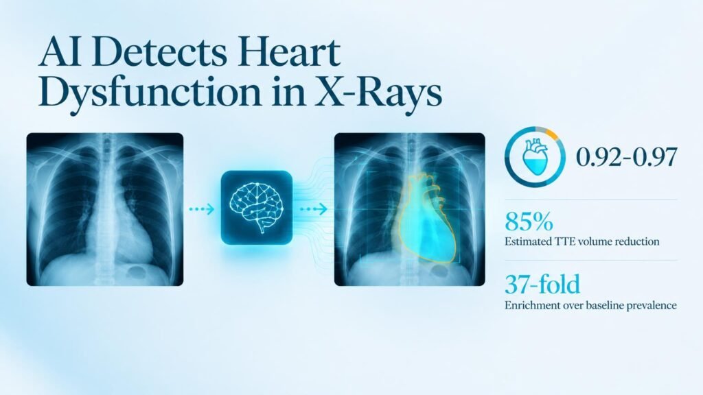
- Reached an area under the curve between 0.92 and 0.97 for severe dysfunction in the two health checkup cohorts.
- Increased detection rates by 87-fold and 37-fold over baseline prevalence in asymptomatic screening populations.
- Maintained low false-negative rates, catching all 12 and 7 patients with ejection fractions under 40% in the checkup groups.
- Estimated an echocardiography volume reduction of up to 85% when used as a screening gatekeeper.
These findings build on broader research examining end-to-end deep learning versus label-based models to identify ventricular dysfunction from routine radiographs. Counterfactual reinforcement learning allowed the tool to map heatmaps directly onto the heart, yielding significantly higher localization precision.
Low Disease Rates Demand Caution
Despite high ROC metrics, the practical deployment of this tool faces a sharp reality check. In screening populations, low disease prevalence pushed the precision-recall area down to 0.04 in both checkup cohorts. High discriminatory power in a trial setting does not eliminate false positives when applied across thousands of healthy individuals.
That low precision means routine implementation would still generate unnecessary secondary scans. Retrospective imaging studies cannot prove that automated screening improves long-term patient outcomes or reduces overall healthcare expenditure.
Health systems should not view these retrospective results as a green light to automate diagnostic pathways. The practical takeaway is to test the model as an optional flag for radiologists rather than an autonomous triage system. Prospective trials must first confirm that cutbacks in echocardiograms do not delay care for patients with borderline ejection fractions.
Read the full study in Radiology: Artificial Intelligence.