A new deep-learning tool dramatically reduces metal artifact errors on internal spinal x-rays, but its failure to replicate those gains on external data shows why clinics cannot yet automate postoperative tracking.
Spinal implants wreck the image quality of postoperative x-rays. The bright metal causes severe streak artifacts that blind standard segmentation algorithms, forcing radiologists to spend valuable time manually measuring spinopelvic parameters. A new framework called RSM, powered by an Artifact Removal Network (ARNAI), attempts to solve this by using autoencoding and inpainting to digitally erase the metal and reconstruct the underlying bone structure. This is essentially digital reconstruction for x-rays.
While the algorithm shines in its home institution, its external validation reveals a familiar, stubborn bottleneck in clinical AI deployment. The software works beautifully on the data it knows, but it falters when introduced to a new clinical environment. This disconnect challenges the assumption that image-clearing algorithms are ready for widespread clinical adoption.
The internal success
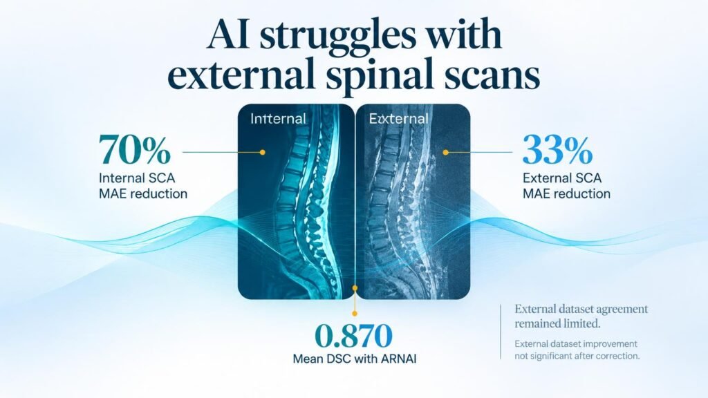
To build and validate the system, researchers retrospectively reviewed lateral lumbar radiographs from two institutions. The internal dataset included **2,486** cases from January 2017 to December 2024. When ARNAI was added to the segmentation pipeline, the overall mean Dice similarity coefficient rose to **0.870** from **0.814**, showing marked improvements at the L3-L5 levels.
Operationally, the tool initially appeared to be a massive time-saver. For implant-containing radiographs in the internal test set, rejected radiographs decreased by **64.21%**, dropping from 95 to 34. The mean absolute error for the L4-L5 segmental Cobb angle plummeted by approximately 70%, dropping to **4.7°** from a baseline of **15.6° to 16.2°**. This improvement was highly significant, even after strict statistical corrections.
The external reality
The narrative shifts when the tool faces data from an outside institution. Testing on an external cohort of **217** patients from October 2021 to September 2025 yielded much weaker results. While the tool managed to reduce rejected radiographs by **65.45%** (from 110 to 38), the actual measurement accuracy did not hold up.
- The L4-L5 segmental Cobb angle error only decreased to a range of **9.5° to 9.7°**, down from **14.2° to 14.5°**.
- This modest 33% reduction failed to remain statistically significant after multiple comparison corrections.
- The final agreement between the AI’s measurements and expert human references remained highly limited.
Why this matters
This performance drop is a major hurdle for clinical utility. A margin of error near 10 degrees on external scans is simply too wide for spinal surgeons, who rely on precise alignment metrics to evaluate fusion success or plan revision surgeries. An algorithm that works well only at its training site cannot be safely deployed across a diverse healthcare network.
Inpainting, by definition, involves guessing what lies beneath an obstruction. When an algorithm guesses incorrectly on an external scan due to variations in imaging equipment or implant types, the clinical consequences can be severe. Miscalculating a Cobb angle could lead to missed hardware failures or unnecessary follow-up imaging.
The study proves that while inpainting networks can clean up metal artifacts on paper, they cannot yet bypass the need for human oversight. If a clinic adopts this software expecting automated postoperative measurements, they risk relying on inaccurate geometry. For now, the tool remains a promising research assistant rather than an autonomous clinical utility, highlighting the massive gap between single-site validation and real-world clinical readiness.
Read the full study in Radiology: Artificial Intelligence.