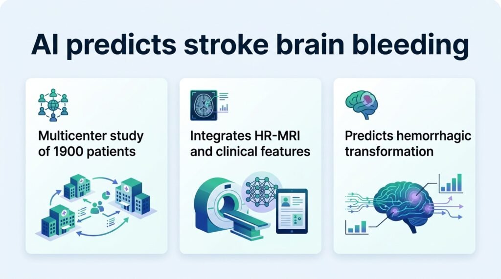
A new machine learning model shows that combining high-resolution MRI with clinical data can spot patients at risk of dangerous brain bleeding before it happens.
When treating an acute ischemic stroke, the biggest fear is hemorrhagic transformation. This is when a blocked vessel begins to bleed, often turning a treatable event into a fatal one. Doctors must decide in minutes whether to dissolve a clot, knowing the treatment itself might trigger this bleeding.
For years, clinicians relied on basic clinical scales to guess this risk. This new study proves that guessing is no longer acceptable. By combining high-resolution magnetic resonance imaging (HR-MRI) with clinical data, researchers built a model that actually maps the biological vulnerability of the brain tissue. This challenges the traditional reliance on simple bedside scores.
The researchers built their model using a massive dataset of 1,400 stroke patients from the First Central Hospital of Baoding. They split this group into a 7:3 ratio for training and testing. To prove the model works in the real world, they also tested it on an independent cohort of 500 patients from Nanjing Gaochun People’s Hospital.
How the model performed
Out of eight machine learning algorithms tested, a random forest model won. It achieved an area under the receiver-operating characteristic curve (AUC) of 0.935 in the training group. More importantly, it maintained a strong AUC of 0.863 in the external validation group, proving it translates well to other hospitals.
- The model identified the hyperintense acute reperfusion marker (HARM) sign as a top predictor.
- Plaque enhancement grade and cerebral microbleeds were critical imaging markers.
- Infarct core volume, baseline NIHSS scores, and fasting blood glucose completed the risk profile.
This is not the first attempt to solve this problem. Previous researchers have tried using post-procedural cone-beam CT images to detect bleeding, as detailed in Neuroradiology. Others have focused on weak lesion feature extraction to make predictions safer. However, this new study shifts the focus to high-resolution vessel wall imaging.
The real-world bottleneck
While the predictive power is impressive, the clinical reality is complicated. HR-MRI scans take time to acquire and analyze. This directly clashes with the “time is brain” rule of acute stroke care, where every minute of delay destroys millions of neurons.
If clinics cannot run these scans rapidly, the model remains a research tool rather than a bedside assistant. The next hurdle is not improving the math, but speeding up the imaging. Until we can acquire high-resolution vessel data in seconds, doctors will still have to make blind choices in the emergency room.
Read the full study in the European Journal of Medical Research.