A new study proves AI can extract three-dimensional genetic data from cheap, flat eye photos, bypassing the need for expensive imaging equipment.
How do you map the genetics of blindness when the necessary 3D eye scans do not exist for millions of patients? For years, researchers have been bottlenecked by the high cost of Optical Coherence Tomography (OCT) devices. This study flips the script by using deep learning to reconstruct 3D retinal structures from simple, flat 2D fundus photographs.
This challenges the assumption that we need expensive, specialized hardware to conduct large-scale genetic mapping. Instead, software can resurrect missing clinical depth from existing, low-tech archives. It shifts the bottleneck of genetic discovery from active data collection to creative computational repurposing.
The flat-to-3D breakthrough
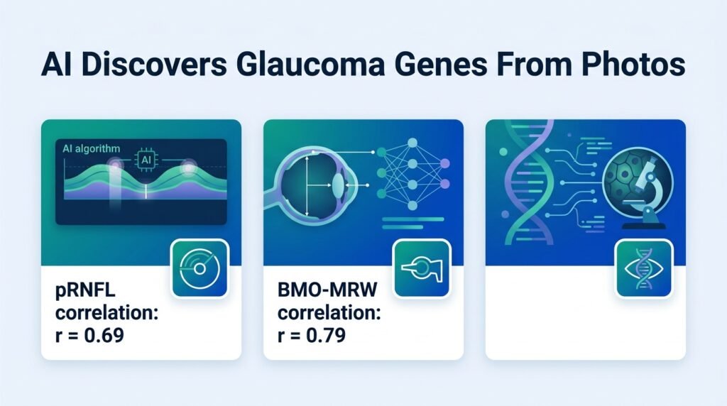
The researchers targeted two critical 3D biomarkers for glaucoma: peripapillary retinal nerve fibre layer (pRNFL) thickness and Bruch’s membrane opening-minimum rim width (BMO-MRW). The AI-derived measurements from 2D fundus images showed strong correlations with actual 3D OCT scans, reaching 0.69 for pRNFL and 0.79 for BMO-MRW. This mathematical bridge allowed the team to analyze cohorts that lacked advanced imaging.
To prove the genetic utility of these virtual measurements, the team ran predictions on the UK Biobank and the Canadian Longitudinal Study on Aging. The genetic correlations between the AI’s predicted shapes and directly measured physical traits were remarkably high, scoring 0.70 for pRNFL and 0.96 for BMO-MRW. This statistical boost allowed the team to identify a massive haul of genetic signals.
Uncovering hidden genetic signals
By expanding the dataset through AI proxies, the study mapped genetic associations at an unprecedented scale. The findings reveal that flat images contain highly specific structural clues that correlate directly with genomic risk factors.
- AI-derived 2D proxies correlated at 0.69 and 0.79 with true 3D scans.
- Genetic correlation reached up to 0.96 for BMO-MRW.
- The method identified 122 loci for BMO-MRW and 29 loci for pRNFL.
- A total of 14 discovered loci are independent of traditional clinical risk factors like vertical cup-to-disc ratio.
This approach exposes a wealth of new biological targets. However, as noted in previous literature on the challenges in automated glaucoma diagnosis, 2D images often suffer from quality variations that can mislead algorithms. Relying on proxies could introduce systemic noise if the underlying training data is biased.
The limits of virtual data
We must remain cautious about the clinical translation of these findings. While a 0.96 genetic correlation is impressive, the initial phenotypic correlation of 0.69 for pRNFL indicates that 2D images still lose structural detail. AI-generated proxies are mathematical estimations, not physical realities.
They can accelerate population-level genetic discovery. Yet they cannot replace physical diagnostic scans in clinical triage where individual accuracy is paramount.
Read the full study in medRxiv.