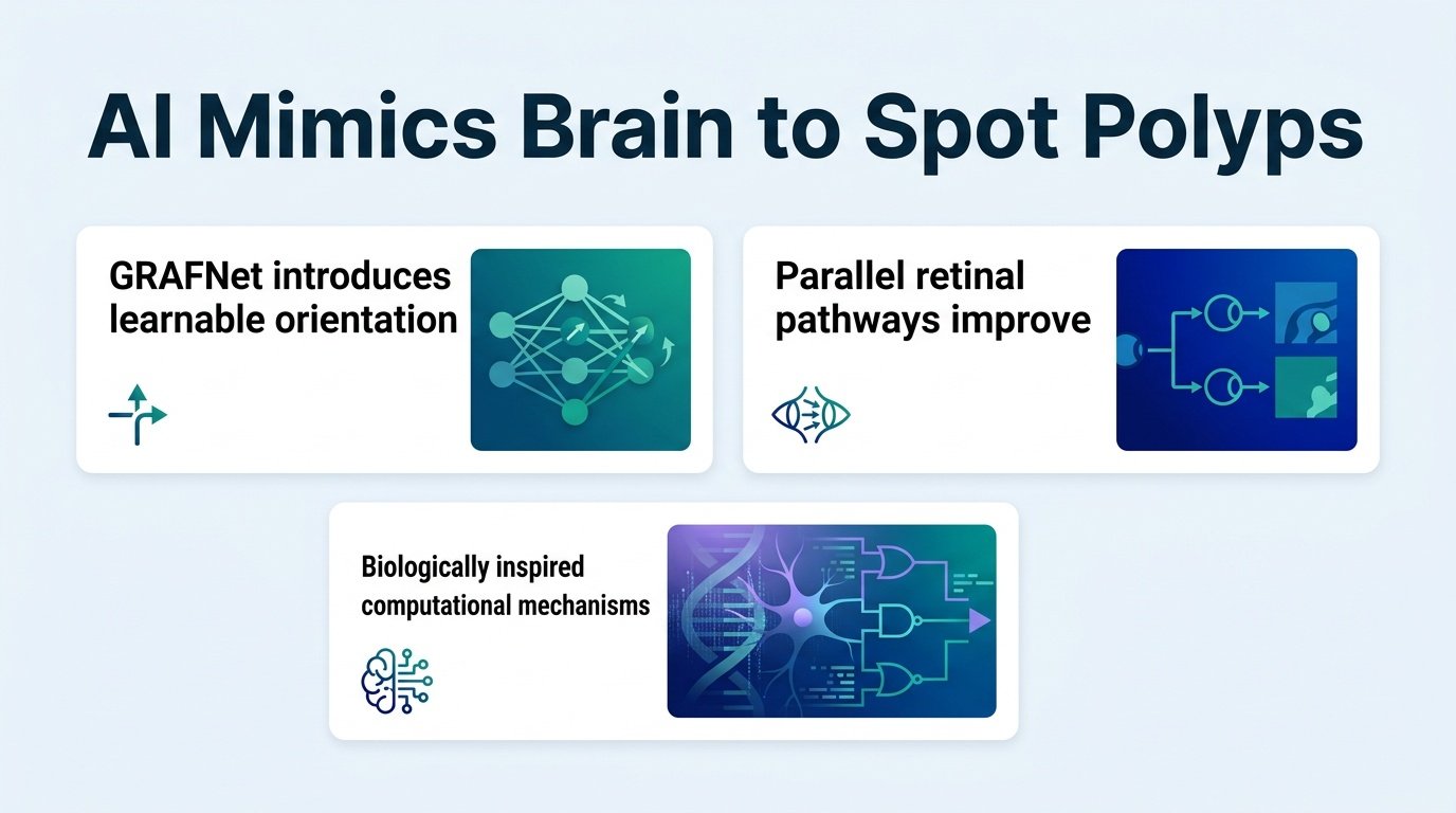
By mimicking how the human brain corrects its own visual mistakes, a new AI model is finding the flat, hidden colon polyps that standard computer vision misses.
Why do state-of-the-art medical AI models still miss flat, subtle polyps during a colonoscopy? The answer lies in a fundamental design flaw: they only look once. Today’s deep learning models are feedforward systems that process an image in a single, irreversible pass. If the first guess is wrong, the system has no way to double-check its work.
This limitation exposes a massive gap between machine vision and human biology. When a human gastroenterologist looks at a blurry screen, their brain does not just register static pixels. It constantly sends feedback down the optic nerve to resolve ambiguity. A new framework called GRAFNet suggests that the path to safer clinical AI is not bigger datasets, but smarter, recurrent architectures that can change their minds mid-run.
To fix this, researchers built three biological mechanisms directly into the neural network. First, they added orientation filters that mimic how the brain’s visual cortex detects edges. Second, they built parallel pathways that compete with each other to suppress background noise. Finally, they introduced predictive coding, which allows the AI to run a feedback loop during inference to refine its predictions.
Traditional computer vision treats image segmentation like a conveyor belt. Pixels go in, a prediction comes out, and the machine moves on. GRAFNet treats it like a conversation. The high-level layers of the network send hypotheses back down to the low-level layers, calculating prediction errors until the image makes sense.
How the model performed
The model showed its real strength on the most difficult visual targets, outperforming standard feedforward networks across several benchmarks:
- A 9.8% improvement in detecting flat lesions compared to the best competing models.
- A 4.8% reduction in misclassifying normal anatomical structures like haustral folds.
- High-accuracy scores including a 0.929 Dice on CVC-ClinicDB, 0.915 on Kvasir-SEG, and 0.946 on CVC-300.
Haustral folds, which are the natural ridges of the colon, frequently trick standard AI into triggering false positives. This forces doctors to waste time investigating normal tissue. GRAFNet’s ability to suppress these competitive features suggests we can finally reduce the alarm fatigue that plagues modern endoscopy suites.
Why feedback loops matter
Flat lesions are the nightmare of colonoscopies. They blend into the colon wall, making them incredibly easy for standard feedforward AI to overlook. By allowing the network to dynamically re-examine ambiguous regions, GRAFNet solves a clinical bottleneck rather than just chasing marginal gains on easy datasets.
But this biological complexity comes with a catch. Running iterative feedback loops during inference takes computational time. In a live colonoscopy, every millisecond of delay matters to an endoscopist. If this closed-loop architecture slows down the video feed, clinicians will not use it, no matter how accurate it is. The next hurdle is proving this brain-like processing can happen in real-time.
Read the full study in Discover Artificial Intelligence.