A new AI ensemble shows impressive accuracy in identifying melanoma, but its reliance on image borders reveals a lingering flaw in computer vision.
Why do highly accurate medical AI models fail when they enter real clinics? The answer usually lies in what the algorithm actually “sees” versus what we think it sees. A model can score near-perfect marks on paper while tracking surgical tape, skin folds, or camera artifacts instead of the actual tumor.
This tension is at the heart of a new study validating an ensemble AI model for skin cancer. By combining two distinct neural networks, researchers built a system with high statistical accuracy. Yet, the real value of this study is not the high score. The real value is the honest look under the hood, which confirms that even with rigorous validation, AI still gets distracted by peripheral noise.
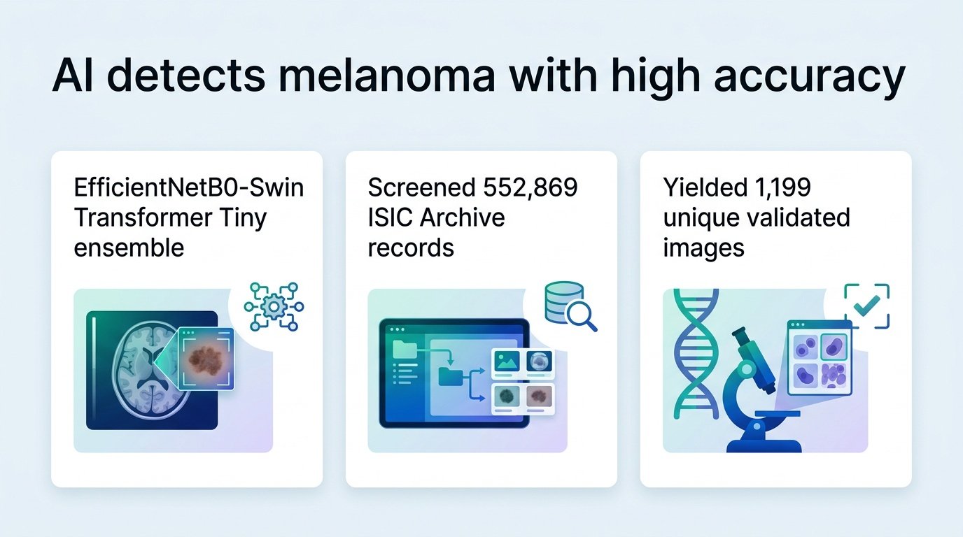
Researchers began by screening 552,869 records from the ISIC Archive. They filtered this massive pool down to 1,199 unique-patient and unique-lesion images, consisting of 578 benign cases and 621 malignant melanomas. Using histopathology as the gold standard, they trained an ensemble combining EfficientNetB0 and Swin Transformer Tiny architectures.
How the model performed
The model achieved strong diagnostic metrics across five-fold validation:
- Mean accuracy reached 0.89325 with a fold-level 95% confidence interval of 0.8538 to 0.9327.
- The area under the receiver operating characteristic curve (ROC-AUC) averaged 0.96348.
- Pooled counts revealed 545 true positives and 526 true negatives.
- The system missed 76 melanomas (false negatives) and flagged 52 benign lesions as cancerous (false positives).
- Overall sensitivity stood at 87.76%, while specificity reached 91.00%.
The peripheral vision problem
These numbers look clean, but the qualitative analysis tells a more complicated story. In two false-positive cases, visual explanations showed the AI activated on peripheral artifacts rather than the lesion itself. When an algorithm decides a mole is cancerous because of a camera lens smudge, clinical trust evaporates.
This highlights a persistent issue in digital dermatology. We are building sharp tools, but we still do not fully control their focus. Furthermore, the researchers used the validation folds for early stopping and checkpoint selection, which can subtly inflate performance. Without external validation on entirely different hospital datasets, these high metrics remain theoretical.
Why this finding matters
A sensitivity of 87.76% means the model still misses more than one in ten melanomas. In a clinical setting, a missed melanoma is a missed window for early intervention. If dermatologists are to rely on this as a safety net, they must know exactly when the AI is looking at the lesion and when it is distracted by the edge of the photo.
Read the full preprint on medRxiv.