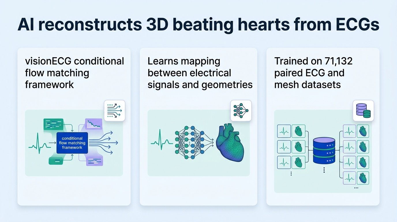
A new AI model reconstructs three-dimensional, moving structures of the human heart using nothing but a standard electrocardiogram.
For decades, clinicians treated electrical signals and physical heart structures as two separate diagnostic tracks. You run an electrocardiogram (ECG) to check the rhythm, but you order an ultrasound or MRI to see the physical muscle. A new framework called visionECG bridges this gap by mapping the relationship between electrical waves and physical cardiac geometries.
That disconnect is the real target here.
This challenges how we think about diagnostic hardware. We usually assume that to get better clinical pictures, we need more expensive machines. This study suggests the data we already collect contains hidden structural dimensions, waiting for the right software to extract them.
Mapping electricity to shape
The system reframes the humble ECG from a simple rhythm strip into a dense, generative map of physical tissue. Built using 71,132 paired ECG and cardiac mesh sequence datasets from the UK Biobank, the model learns how electrical patterns dictate physical movement. It reconstructs a moving, three-dimensional representation of the left ventricle using only an ECG and basic patient demographics.
To prove this was not just a neat trick trained on clean data, researchers tested the model on 5,000 external patients with ECG-echocardiogram pairs. The AI successfully flagged structural abnormalities and visualised functional defects. It allowed researchers to calculate both global and regional heart parameters without placing an imaging probe on the patient’s chest.
This means a simple clinic visit could soon yield insights previously restricted to a specialist imaging referral.
The limits of prediction
The implications go beyond saving money on imaging equipment. By translating low-dimensional signals into high-dimensional physical models, clinicians can track regional heart wall motion issues in real time. This shifts the ECG from a passive monitoring tool to an active, generative diagnostic asset that could democratise cardiac screening in rural clinics.
However, a synthetic heart is still a prediction, not a direct observation. The model relies on probabilistic mapping, meaning it infers structure based on historical patterns rather than seeing the patient’s unique physical anomalies in real time. If a patient has an unusual structural defect that does not correlate with typical electrical changes, the model might miss it entirely.
We must not mistake statistical inference for actual clinical sight. While this tool can screen for hidden risks, it cannot replace the ground truth of a physical scan when surgery is on the line. It serves as an advanced triage mechanism, not a final diagnostic word.
- Processed 71,132 paired datasets to map electrical signals to physical heart shapes.
- Validated on 5,000 external patients using real-world ultrasound data.
- Reconstructed moving 3D left ventricles using only basic patient demographics and ECGs.
Read the full preprint study on medRxiv.