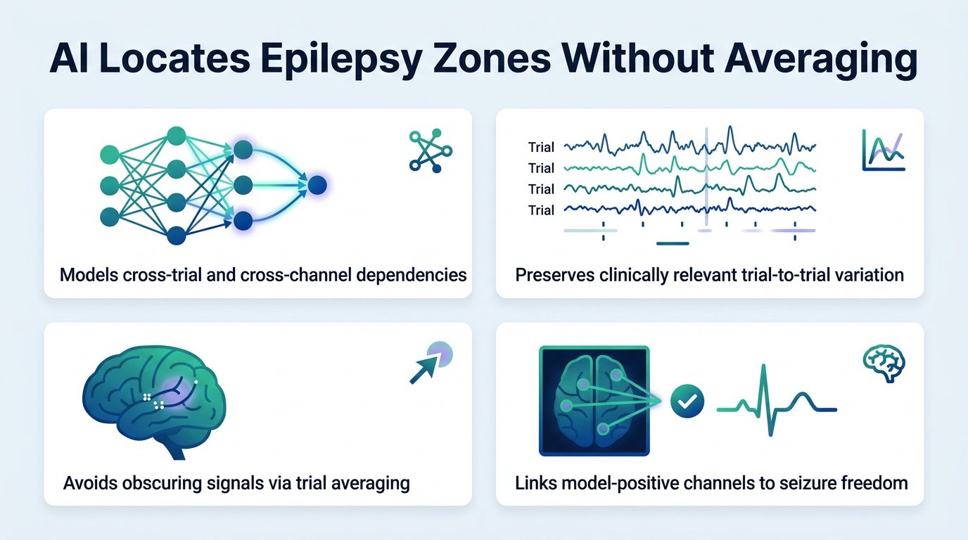
A new deep learning model pinpoints seizure-generating brain tissue by analyzing individual electrical pulses rather than averaging them together.
For decades, neurologists have smoothed out brain signal noise by averaging repeated electrical stimulation trials. This mathematical shortcut cleans up the clinical data. However, it likely throws away the very variations that signal where a seizure starts.
It treats fluctuation as a nuisance rather than a diagnostic clue.
This study challenges the dogma of signal averaging. By proving that trial-to-trial variation is valuable signal rather than useless noise, it suggests we have been throwing away critical diagnostic data. This shift in perspective could redefine how clinicians map the brain before epilepsy surgery.
The practical implications are immediate. If clinical software no longer needs dozens of repeated stimulations to get a clean reading, diagnostic sessions can become much shorter. This reduces the physical burden on patients undergoing invasive brain monitoring.
Ditching the average
Researchers developed a Hierarchical Attention Transformer (HAT) to analyze single-pulse electrical stimulation (SPES) data. The model was tested on data from 35 patients using patient-held-out, repeated five-fold cross-validation. Instead of flattening the data, the model tracks how responses shift across channels and individual trials.
The results show that preserving these raw, messy variations yields a much clearer picture of the brain. The key findings include:
- The HAT model achieved a seizure onset zone concordance AUROC of 0.762, significantly outperforming the trial-averaged baseline of 0.721.
- The model maintained this high performance even when restricted to just 1 or 5 trials during testing.
- The mean paired difference in performance between the models was 0.041, confirming a statistically meaningful advantage with a Holm-adjusted p-value of 0.0197.
The clinical reality check
Despite the improved mapping, the study hit a major roadblock when predicting actual patient recovery. The proportion of model-positive brain channels resected did not reliably predict whether a patient would remain seizure-free after surgery. This association was weak, yielding an AUROC of just 0.634 with a non-significant p-value of 0.102.
This gap highlights a persistent problem in clinical AI. The model was trained to find the “seizure onset zone” as defined by human doctors, not to predict surgical success. If the human definition of the seizure zone is imperfect, an AI trained on that definition will simply replicate those human limitations.
Future work must train these models directly on patient outcomes rather than clinical proxies. Until then, this technology remains a sharper mapping tool, but not yet a guarantee of a cure.
Read the full study in medRxiv.