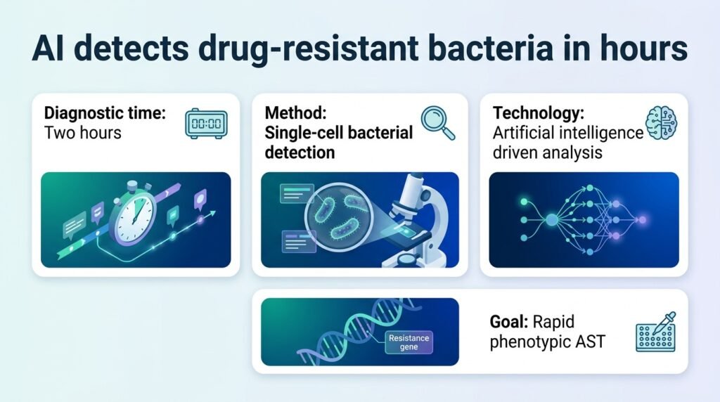
A new microfluidic AI platform cuts bacterial resistance testing down to two hours, challenging the slow timelines of traditional lab cultures.
Why does it still take two days to find out if an antibiotic will save a patient with sepsis? Doctors routinely prescribe broad-spectrum drugs blindly while waiting 24 to 48 hours for standard lab cultures to grow. This delay fuels the rise of superbugs and leaves patient survival to chance.
This new research challenges the accepted compromise of empirical therapy. By merging microfluidics with computer vision, the system shifts the diagnostic bottleneck from biological growth to computational speed. It proves that we do not need to wait for massive bacterial colonies to develop to see if a drug works. We only need to watch a single cell’s immediate physical reaction.
Watching single cells
The platform captures time-lapse images of individual bacterial cells under controlled antibiotic exposure. A specialized U-Net AI model automatically segments and tracks how these single cells respond to drugs. This bypasses the traditional requirement of growing millions of cells to get a visible signal.
In testing, the AI successfully identified 96% of individual Escherichia coli (E. coli) cells. Crucially, the model recorded zero false-positive predictions on bacteria-free images, a vital metric for avoiding clinical misdiagnosis.
Two hours to profile
The speed of this approach redefines clinical timelines. The system quantified how E. coli responded to different doses of ciprofloxacin and trimethoprim/sulfamethoxazole. It delivered complete resistance profiles and minimum inhibitory concentrations (MICs) within just two hours.
These rapid results matched the accuracy of standard broth microdilution, the slow-but-steady benchmark of clinical microbiology. Similar rapid imaging approaches, such as those evaluated in this 2026 performance evaluation, confirm that single-cell imaging is a viable path to faster phenotypic testing.
- Identified 96% of single E. coli cells.
- Achieved zero false-positives on clean, bacteria-free images.
- Delivered drug resistance profiles and MICs within two hours.
- Successfully isolated and identified E. coli and S. aureus in complex blood matrices.
The clinical reality check
Despite the impressive speed, implementation hurdles remain. The study successfully tested the platform on E. coli and Staphylococcus aureus (S. aureus) inside complex blood matrices. However, blood is a messy medium, and scaling this to handle polymicrobial infections or rare pathogens will require much larger training datasets.
Furthermore, clinical labs are notoriously slow to adopt complex microfluidic hardware. For this technology to succeed outside the research lab, manufacturers must package these delicate fluid channels into cheap, fool-proof cartridges. Until then, the two-hour diagnostic remains a promise rather than a bedside reality.
Read more about this research in Advanced Science.