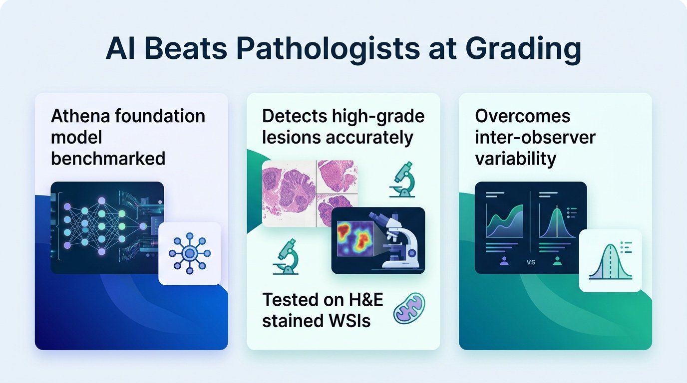
A new study shows that AI foundation models can standardize cervical cancer screening across different countries, outperforming human pathologists where they struggle most.
Pathology has a consistency problem. When different doctors look at the same cervical biopsy, they often disagree on whether a lesion is high-grade or low-grade. This subjectivity means patients in one clinic might receive aggressive surgery, while identical cases elsewhere are merely watched.
A new pre-print challenges the assumption that AI cannot handle this real-world variability. By testing models across diverse healthcare systems, researchers proved that algorithms can bring much-needed standardization to a highly subjective field.
Testing across five nations
To prove the AI could work globally, researchers gathered H&E whole-slide images from five distinct regions: Portugal, Cambodia, Germany, Poland, and Scotland. They benchmarked their custom pipeline, called Athena, against four prominent pathology foundation models: H-optimus-0, Hibou-L, Midnight-12k, and Virchow.
Athena led the pack. It achieved a mean area under the curve (AUC) of 0.931 and demonstrated remarkable stability, with a cross-country standard deviation of just 0.022. This consistency is crucial because medical imaging hardware and staining techniques vary wildly between international labs.
AI versus human eyes
The real test came when comparing the AI directly to trained pathologists using a gold-standard, p16-confirmed dataset. The algorithm significantly closed the gap on missed cases.
- The model raised diagnostic sensitivity to 95%, up from the pathologists’ 84%.
- Specificity remained nearly identical, with the model scoring 85% compared to humans at 84%.
- Model errors were tightly clustered at the tricky boundary between low-grade and high-grade lesions.
- Pathologists’ errors were scattered across a much broader range of misclassifications.
This performance gap matters because human error in pathology is often random, driven by fatigue or subjective bias. The model, by contrast, only stumbles where the biology itself is genuinely ambiguous. This predictable failure mode makes the AI a highly reliable safety net.
This work builds on previous efforts to automate women’s health screenings, such as UniCAS, which focused on cervical cytology. Moving from cytology to tissue biopsies is a major step forward for clinical workflows. It suggests foundation models are mature enough to handle complex tissue architecture, not just isolated cells.
The limits of the data
We must acknowledge the study’s boundaries. These findings come from a pre-print, and the pipeline still requires validation in active, prospective clinical trials before replacing human eyes. Furthermore, while the model excels at binary classification, clinical reality is rarely binary.
If these models can be successfully integrated into lab software, they will not replace pathologists. Instead, they will act as an unblinking second reader, catching the 11% of high-grade lesions that humans currently overlook.
Read the full study on medRxiv.