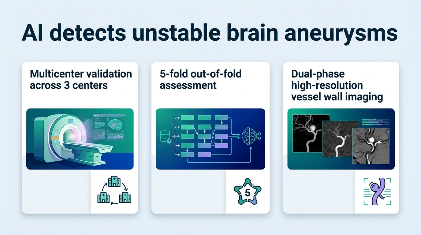
A new deep learning model successfully flags unstable brain aneurysms across different hospitals, proving that AI can read subtle blood vessel walls without losing accuracy outside its training ground.
Radiologists face a high-stakes guessing game when evaluating silent brain aneurysms. A rupture is catastrophic, yet unnecessary brain surgery carries its own grave risks. Deciding which bulges are unstable requires reading microscopic changes on a blood vessel wall, a task that is highly subjective and prone to human error.
This is where standard imaging algorithms usually fail. They excel in the lab where they are trained, but crumble when deployed at a different hospital with different scanners. This study challenges that limitation, proving that a wall-focused AI can maintain its diagnostic accuracy across independent medical centers.
Consistency across different scanners
Researchers built a dual-phase framework called WCE-Net combined with a transformer-based branch to analyze high-resolution vessel wall scans. By fusing non-contrast and contrast-enhanced imaging, the model captures both the physical shape of the wall and how tissue behaves under contrast. They trained and tested the system on a dataset of 629 patients with 773 aneurysms across three different medical centers. Center 1 served as the development hub, while Centers 2 and 3 tested how well the AI performed on entirely unfamiliar patient data.
The AI did not just work in its home environment. It achieved an area under the curve (AUC) of 0.908 at the development center, and held strong with AUCs of 0.857 and 0.855 at the two external validation sites. Its Brier scores, which measure how close predicted probabilities are to actual outcomes, remained tight at 0.119, 0.153, and 0.150 respectively.
- An internal accuracy AUC of 0.908 that only slightly dipped to 0.855 in external testing.
- Low Brier scores across all centers, indicating highly calibrated probability estimates.
- Visual heatmaps that partially overlapped with actual high-signal vessel wall regions.
The trust problem remains
This consistency is a major step forward, but the trust gap in clinical AI still lingers. The researchers used 3D heatmaps to show where the AI was looking, finding only partial spatial overlap with the high-signal regions human radiologists typically flag. This disconnect highlights a broader trend in the field, as discussed in a recent review on deep learning in vascular wall imaging. If the AI is seeing risk factors that humans cannot define, clinicians may hesitate to trust its recommendations.
We must also note the study’s retrospective design. Real-world clinical workflows are messy, and retrospective data often hides confounding variables. Until this tool is tested prospectively in live clinical decision-making, it remains a highly promising proof of concept rather than a tool ready for daily practice.
This analysis is based on a study published in medRxiv.