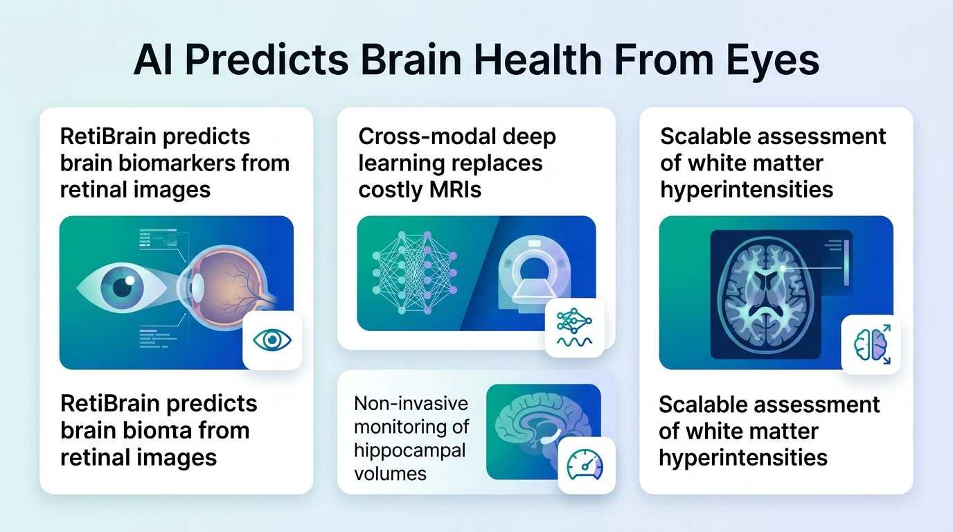
A new deep learning model bypasses expensive brain scans by reading signs of dementia and stroke directly from the back of the eye.
Can a simple photo of your retina replace a multi-million dollar MRI machine? For years, researchers have known the eye is a window to the brain, but translating eye blood vessels into precise brain measurements has been a guessing game.
This new model, RetiBrain, challenges the assumption that we need actual brain imaging to track cognitive decline. By training an AI to translate eye photos into simulated MRI metrics, it bridges the gap between cheap screening and expensive neurology.
This shift forces us to rethink how we define a biomarker. If a proxy measurement is accurate enough to predict disease, do we really need the master image?
This is not just about cheaper diagnostics. It means we can track white matter hyperintensities and hippocampal shrinkage—the physical footprints of dementia—years before symptoms show up, using equipment already sitting in every local optometrist’s office. If we can spot hippocampal atrophy through a routine eye exam, we can enroll patients in clinical trials long before their cognitive function begins to fail.
Mapping the eye
The researchers built RetiBrain by training a cross-modal deep learning framework on paired eye photos and brain MRIs. It targets six specific biomarkers linked to white matter hyperintensities and hippocampal volume.
In head-to-head testing, RetiBrain easily beat RETFound, a leading retinal foundation model. It boosted the mean Pearson correlation coefficient by 0.309, jumping from 0.240 to 0.549. For periventricular white matter hyperintensities, the correlation reached 0.640.
Tracking long-term risk
The real test of any biomarker is whether it can predict actual sickness over time. Researchers validated the model using a massive longitudinal dataset to see if these eye-derived brain metrics translated to real-world diagnoses.
- The validation cohort tracked 2,082 participants and analyzed 4,164 retinal images.
- The study followed these patients for up to 15 years.
- RetiBrain predicted dementia with an AUROC of 0.824.
- Each standard deviation increase in predicted risk multiplied the hazard ratio by 2.500 (95% CI: 2.201-2.840).
We must look at these numbers with a critical eye. A Pearson correlation of 0.549 means the AI is still a proxy, not a perfect replacement for a physical MRI. It is an estimation tool, and clinical decisions will still require actual neuroimaging to confirm structural damage. We also do not know how this model performs across diverse ethnic populations, as retinal pigmentation can affect image quality.
Furthermore, these findings come from a preprint that has not yet undergone formal peer review. However, if these results hold up, they suggest we can shift brain health monitoring from reactive crisis management to proactive, routine eye exams. Doctors could catch vascular brain injuries before a patient ever has a stroke.
Read the full study in medRxiv.