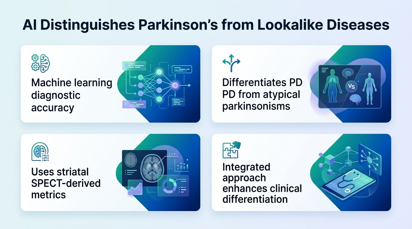
A new machine learning approach shows that while AI can easily spot the difference between Parkinson’s and its deadlier lookalikes, pinpointing the exact disease remains incredibly difficult.
How do you treat a patient when their brain scans look almost identical to three other fatal conditions? For years, clinicians have struggled to differentiate Parkinson’s disease from atypical parkinsonian syndromes in their early stages. The stakes are incredibly high because these lookalikes progress much faster and do not respond to standard Parkinson’s drugs.
A new study tackles this diagnostic bottleneck. By combining advanced SPECT brain imaging with a random forest algorithm, researchers attempted to map the precise boundaries of these diseases. The analysis reveals a stark disconnect that should make clinicians pause.
The AI achieved a stellar area under the curve (AUC) of up to 0.95 when simply separating Parkinson’s from atypical syndromes. Yet, when tasked with classifying the exact subtype of disease, the algorithm’s overall accuracy plummeted to 64%. This gap tells us that while binary sorting is nearly solved, fine-grained multi-class diagnosis still evades our best algorithms.
Mapping the brain’s pathways
The study analyzed a large cohort of 706 patients. This included 487 individuals with Parkinson’s disease and 219 with atypical parkinsonisms. Within the atypical group, researchers looked at 127 patients with progressive supranuclear palsy, 37 with multiple system atrophy parkinsonian type, 12 with multiple system atrophy cerebellar type, and 43 with corticobasal degeneration.
Every patient underwent a [¹²³I]FP-CIT SPECT scan to measure dopamine transporters in the brain. Instead of just looking at raw images, the team calculated specific binding ratios across anatomical regions like the caudate and putamen, alongside functional zones like sensorimotor and limbic networks. This dense dataset was then fed into a random forest classifier.
Where the algorithm stumbles
The machine learning model mapped two distinct diagnostic pathways to sort the patients. The key findings from this automated sorting include:
- The caudate-to-putamen and sensorimotor-to-limbic ratios were the strongest differentiators, yielding an AUC of up to 0.95.
- The random forest model achieved an overall multiclass accuracy of only 64%.
- Per-class specificities remained high, exceeding 84% across the different conditions.
- A lower caudate-to-posterior putamen ratio successfully grouped progressive supranuclear palsy, corticobasal degeneration, and multiple system atrophy cerebellar type.
- A higher ratio instead grouped Parkinson’s and multiple system atrophy parkinsonian type, using ipsilateral caudate uptake to help separate them.
The diagnostic reality check
This finding forces us to rethink the role of AI in neurology clinics. A 64% accuracy rate means the algorithm cannot replace clinical judgment for specific subtyping. However, the high specificity of over 84% means the tool is excellent at ruling diseases out, preventing misdiagnosis and sparing patients from ineffective treatments.
The study is limited by its cross-sectional design, meaning it captures a single snapshot in time. To truly validate these diagnostic pathways, we need longitudinal data tracking how these algorithmic predictions hold up as the diseases progress.
Read the full study in the European Journal of Nuclear Medicine and Molecular Imaging.