A new open-source AI model successfully automates the tedious manual tracking of heart chambers and blood flow velocity, moving cardiac imaging past simple ventricular checks.
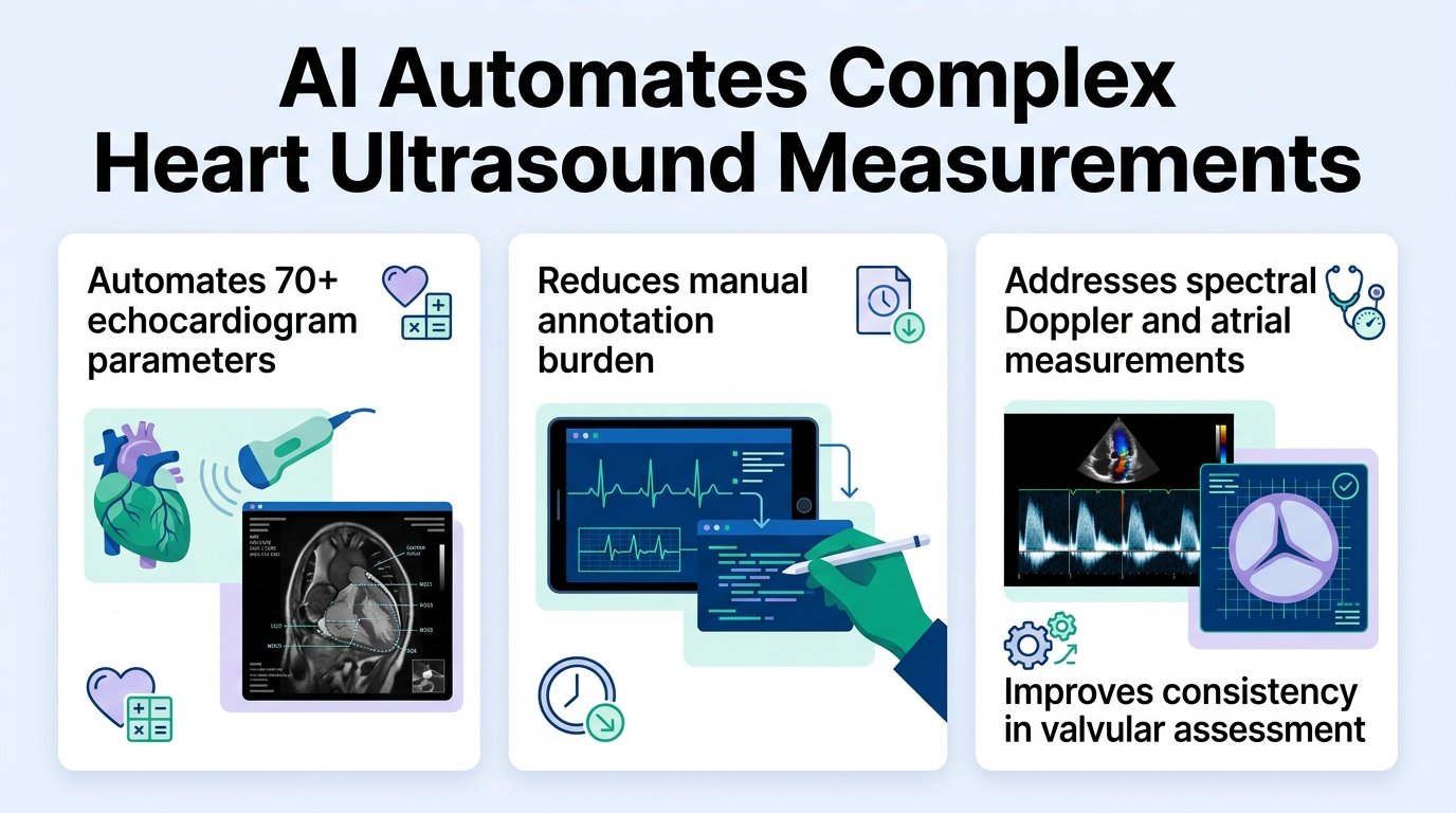
Why does cardiac AI only look at the easy parts of the heart? For years, developers have focused heavily on the left ventricle, leaving clinicians to manually measure atrial sizes and complex Doppler waves. This manual burden slows down clinics and introduces massive human error.
A new study introduces EchoNet-Segmentation to tackle these ignored parameters. This shift challenges the current obsession with massive, generalist medical AI. By focusing on highly specific tasks, the researchers prove that targeted models can outperform broad foundation models where it actually matters.
The researchers built and trained their models using a massive dataset of 186,712 sonographer-annotated images from 93,978 studies representing 56,855 patients at Cedars-Sinai Medical Center. They did not just test the tool in a clean lab. They validated it across a temporal split, an external Kaiser Permanente Northern California cohort, and the public MIMIC-Echo dataset.
How the AI performed
- On the internal test set, the AI achieved an R2 of 0.817 to 0.882 for velocity-time integral (VTI) measurements, with a mean absolute error (MAE) of 1.13 to 3.80 cm.
- For left and right atrial area segmentation, the model hit an R2 of 0.675 to 0.747 with an MAE of 2.48 to 2.52 cm2.
- In the external Kaiser Permanente cohort, performance remained strong, showing an R2 of 0.575 to 0.859 for VTI and 0.803 to 0.876 for atrial area.
These results show a high level of generalizability across different hospital systems and ultrasound vendors. While previous efforts like the ventricular segmentation algorithm based on transfer learning and GAN focused heavily on the ventricles, expanding to atrial and Doppler measurements has been notoriously difficult. The new model’s ability to maintain accuracy on external datasets addresses a major hurdle highlighted in recent multi-centric evaluations of AI generalization.
Why this matters
This is not about saving a few minutes per scan. Automating VTI and atrial measurements means clinics can finally standardize diastolic and valvular heart disease assessments. Human sonographers often disagree on where to draw these boundaries, but a consistent AI baseline reduces this variability.
Furthermore, the model outperformed a prominent open-source medical image foundation model using prompt configurations. This is a critical reality check for the industry. It suggests that specialized, task-specific architectures remain superior to generalist “all-in-one” medical models for precise clinical quantification.
The limits of the data
There are still clear boundaries to these findings. The external validation cohorts, while diverse, still rely on retrospective data. We need prospective, real-time clinical trials to see if this automation actually reduces clinician burnout or improves patient outcomes in busy diagnostic pipelines.
The source study was published in medRxiv.