High-tech cancer diagnostics are currently a luxury of the wealthiest health systems, but a shift in how we analyze basic tissue slides could soon level the playing field.
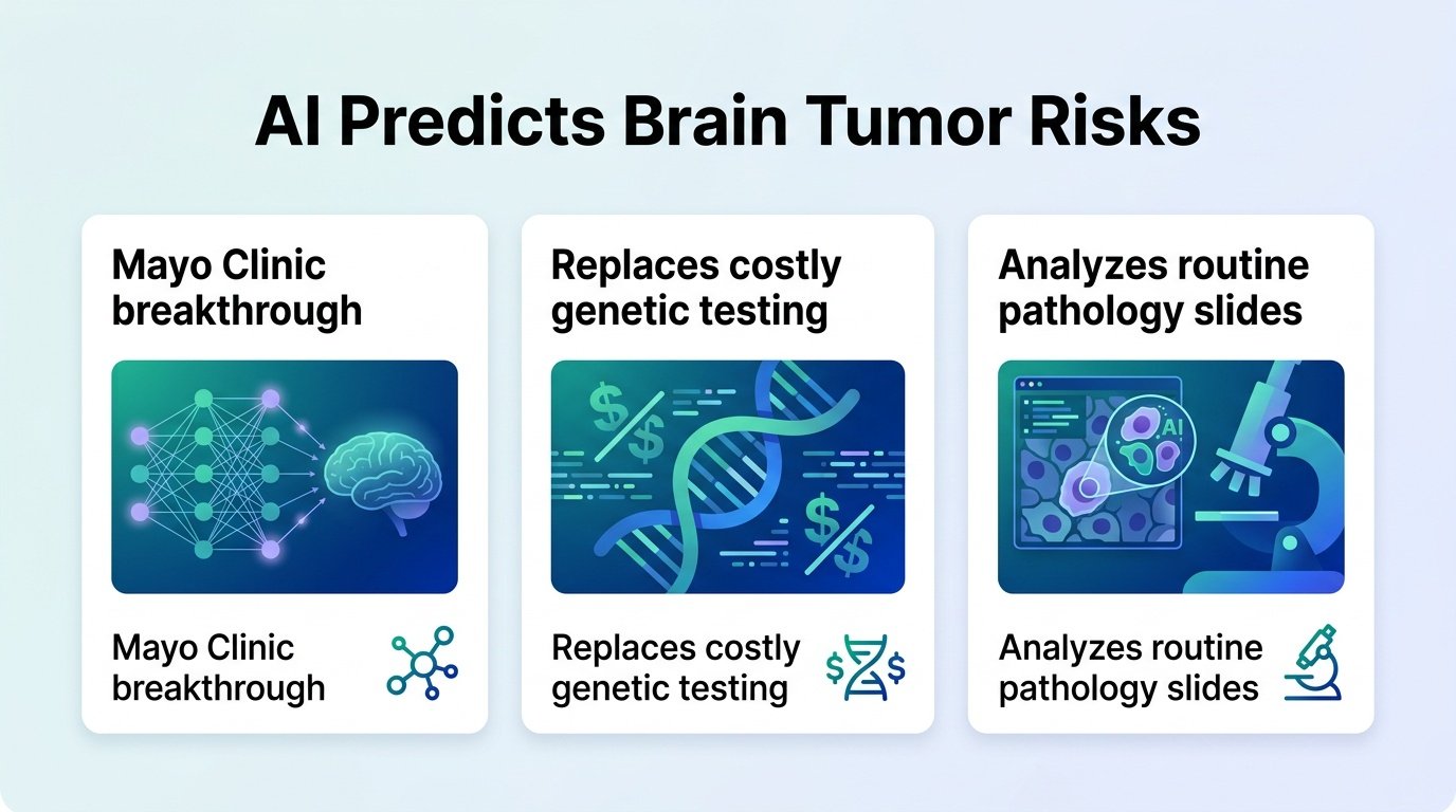
Precision oncology has a bottleneck. To predict if a brain tumor like a meningioma will return, doctors increasingly rely on DNA methylation profiling. This advanced genetic test is expensive and requires specialized infrastructure. Most patients worldwide simply cannot access it.
What if the answers are already hiding in plain sight?
The Digital Shortcut
Researchers trained a deep learning model on routine pathology slides from 672 patients. Instead of sequencing DNA, the AI analyzed standard tissue images to extract molecular insights and predict tumor recurrence.
This approach turns a simple microscope slide into a rich data source. By recognizing subtle visual patterns invisible to the human eye, the algorithm maps out the tumor’s future behavior. The AI’s risk predictions remained highly accurate even when accounting for traditional factors like patient age and surgical success, effectively bypassing the need for costly genetic sequencing.
The Reality Check
This is not just about saving money. It is about democratization. If standard slides can yield molecular-level insights, advanced cancer prognosis moves from elite academic centers to community hospitals.
However, a critical hurdle remains. This analysis was retrospective. Different labs use different staining techniques and scanners, and an AI trained on one hospital’s data often stumbles when deployed elsewhere. Before clinicians can trust an algorithm to guide post-surgery monitoring, these models must prove their accuracy in prospective, real-world trials across diverse hospital systems.