⚡ Quick Summary
This study evaluated the clinical utility of an FDA-cleared AI tool, Neosoma HGG, for automated tumor volume segmentation in patients with high-grade gliomas (HGGs). The findings indicate that while AI significantly reduces segmentation time, there are notable limitations in distinguishing between tumor progression and pseudo-progression.
🔍 Key Details
- 📊 Dataset: 214 subjects from public datasets and 49 from an institutional cohort
- 🧩 Features used: MRI sequences, including contrast-enhancing (CE) and T2-FLAIR volumes
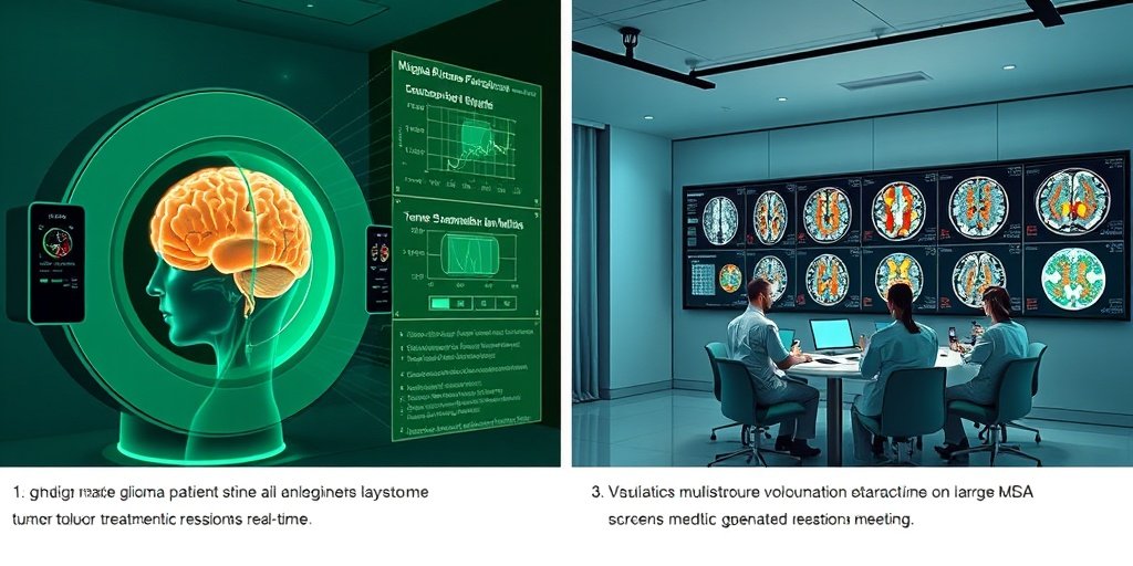
- ⚙️ Technology: Neosoma HGG AI tool for tumor volume segmentation
- ⏱️ Segmentation time: Reduced from 210.5s (pre-operative) and 179s (post-operative) to just 15s (AI)
🔑 Key Takeaways
- 🤖 AI-based segmentation offers a significant reduction in time for tumor volume assessments.
- 📈 Moderate agreement (k = 0.45) with multidisciplinary tumor board (MDTB) assessments for progressive disease.
- ⚠️ Limitations in distinguishing pseudo-progression from true tumor progression were noted.
- 🔄 T2-FLAIR volumes showed significant variability across AI platforms.
- 🌟 Neosoma HGG demonstrates potential for improving efficiency in monitoring HGG.
- 📉 Further refinement is needed to enhance specificity and integrate multimodal imaging data.
- 🏥 Study conducted across multiple datasets, including institutional and public sources.
📚 Background
High-grade gliomas (HGGs) are aggressive brain tumors that necessitate continuous imaging for effective treatment management. Traditionally, this process has relied on manual MRI measurements, which can be both labor-intensive and inconsistent. The introduction of artificial intelligence (AI) tools, such as Neosoma HGG, promises to streamline this process, yet their practical application in clinical settings remains underexplored.
🗒️ Study
This study aimed to validate the AI-based tool Neosoma HGG for quantifying tumor volumes in patients with HGG. Researchers analyzed a total of 1648 MRI sequences from 95 patients across three datasets, comparing AI-derived volumes with those obtained through expert manual assessments and other AI platforms.
📈 Results
The results revealed that Neosoma HGG significantly reduced segmentation time to just 15 seconds, compared to traditional methods that took over 200 seconds. However, the AI tool showed only a moderate agreement with MDTB assessments, particularly struggling to differentiate between pseudo-progression and actual tumor progression. Additionally, T2-FLAIR-derived volumes exhibited significant discrepancies across different AI platforms.
🌍 Impact and Implications
The findings from this study highlight the potential of AI-based volumetric segmentation to enhance the efficiency and standardization of monitoring HGG. However, the moderate concordance with MDTB assessments and challenges with FLAIR imaging underscore the need for further refinement. As AI continues to evolve, it could serve as a valuable clinical decision support tool, ultimately improving patient outcomes in neuro-oncology.
🔮 Conclusion
This study underscores the transformative potential of AI in the assessment of high-grade gliomas. While Neosoma HGG demonstrates significant advantages in terms of efficiency, its current limitations must be addressed to fully realize its clinical utility. Continued research and development in this area are essential for integrating AI into routine clinical practice, paving the way for improved patient care.
💬 Your comments
What are your thoughts on the integration of AI in monitoring high-grade gliomas? We would love to hear your insights! 💬 Share your comments below or connect with us on social media:
Artificial intelligence-based volumetric measurements for longitudinal clinical assessment of treatment response in high-grade gliomas: Validation across institutional and public datasets.
Abstract
BACKGROUND: High-grade gliomas (HGGs) require ongoing imaging to guide treatment, traditionally relying on labor-intensive and variable manual MRI measurements. While FDA-cleared artificial intelligence (AI) tools offer automated tumor volume segmentation, their clinical utility in decision-making remains understudied. This study assesses the utility and limitations of an FDA-cleared AI-based tool across public and institutional datasets, comparing its output with multidisciplinary tumor board (MDTB) assessments.
METHODS: We applied the FDA-cleared, AI-based tool Neosoma HGG to quantify tumor volumes in 214 subjects from public datasets and 49 from an institutional cohort. AI-derived volumes were compared to expert manual and other AI-based measurements. Therapeutic response assessments using RANO criteria were evaluated against MDTB diagnoses. Segmentation times were analyzed using mixed-model regression.
RESULTS: We analyzed 1648 MRI sequences of 95 HGG patients across three datasets. Contrast-enhancing (CE) tumor volumes were consistent across AI platforms, and Neosoma HGG significantly reduced segmentation time (pre-operative: 210.5s, post-operative: 179s vs. 15 s, P < .0001). AI-informed disease state assessments showed an overall moderate agreement with MDTB diagnoses for progressive disease (k = 0.45, P < .00001). Key discrepancies arose from limitation of Neosoma HGG in distinguishing pseudo-progression from tumor progression. T2-FLAIR-derived volumes varied significantly between AI platforms (P < .001), with discordances largely due to over-segmentation beyond the tumor region.
CONCLUSION: AI-based volumetric segmentation has the potential to improve efficiency and standardization in monitoring HGG, especially for CE tumor burden. However, moderate concordance with MDTB assessments and difficulties with FLAIR imaging underscore its current limitations. AI should serve as a clinical decision support tool, with further refinement needed to improve specificity and integrate multimodal imaging data.
Author: [‘Asfaw ZK’, ‘Young T’, ‘Hernandez Marquez G’, ‘Brown C’, ‘Tomalin LE’, ‘Belani P’, ‘Doshi A’, ‘Germano IM’]
Journal: Neurooncol Adv
Citation: Asfaw ZK, et al. Artificial intelligence-based volumetric measurements for longitudinal clinical assessment of treatment response in high-grade gliomas: Validation across institutional and public datasets. Artificial intelligence-based volumetric measurements for longitudinal clinical assessment of treatment response in high-grade gliomas: Validation across institutional and public datasets. 2026; 8:vdag045. doi: 10.1093/noajnl/vdag045