⚡ Quick Summary
This study developed ultrasound-based machine learning models to enhance the non-invasive differentiation between Sclerosing Adenosis (SA) and Invasive Ductal Carcinoma (IDC). The models demonstrated impressive performance, achieving an AUC of 0.9758 for the ROI-level model and 0.9653 for the patient-level model, indicating their potential in clinical diagnostics.
🔍 Key Details
- 📊 Dataset: 2046 ultrasound images from 772 patients
- 🧩 Features used: 18 features for ROI-level model, 9 features for patient-level model
- ⚙️ Technology: XGBoost for ROI-level, Logistic Regression for patient-level
- 🏆 Performance: AUC of 0.9758 (ROI-level) and 0.9653 (patient-level)
🔑 Key Takeaways
- 🔍 Non-invasive differentiation between SA and IDC is crucial for accurate diagnosis.
- 🤖 Machine learning models were developed using ultrasound images, showcasing their potential in diagnostics.
- 🏆 XGBoost achieved an AUC of 0.9758 in the test cohort for the ROI-level model.
- 📈 Logistic regression reached an AUC of 0.9653 for the patient-level model.
- 💡 Feature importance was highlighted, with “Original shape Major Axis Length” being critical for identifying IDC.
- 🌍 External validation confirmed the robustness of the models across different datasets.
- 🛠️ Explainability was enhanced through SHAP, allowing for better understanding of model decisions.
- 🩺 Potential impact includes reducing misdiagnoses and improving early detection of IDC.
📚 Background
The differentiation between Sclerosing Adenosis (SA) and Invasive Ductal Carcinoma (IDC) has long posed a challenge in breast cancer diagnostics. Traditional methods often rely on invasive procedures, which can lead to unnecessary stress and complications for patients. The integration of machine learning with ultrasound imaging presents a promising avenue for enhancing diagnostic accuracy while maintaining a non-invasive approach.
🗒️ Study
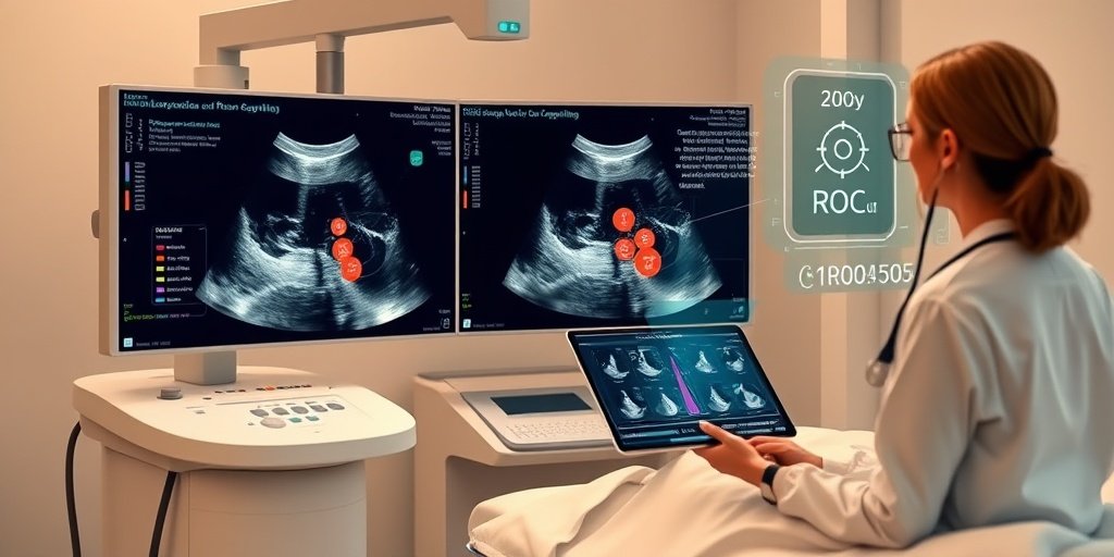
This study aimed to develop machine learning models that leverage breast ultrasound images to facilitate the non-invasive differentiation between SA and IDC. A total of 2046 ultrasound images were collected from 772 patients, with Regions of Interest (ROI) delineated for feature extraction. The dataset was meticulously split into training and test cohorts, and various classifiers were employed to optimize model performance.
📈 Results
The results were promising, with the XGBoost model achieving an impressive AUC of 0.9758 in the test cohort for the ROI-level model, and 0.9906 in the validation cohort. For the patient-level model, logistic regression yielded an AUC of 0.9653 in the test cohort and 0.9846 in the validation cohort. The feature “Original shape Major Axis Length” emerged as the most significant predictor for IDC, indicating its potential utility in clinical settings.
🌍 Impact and Implications
The implications of this study are significant. By providing a non-invasive tool for differentiating between SA and IDC, these machine learning models could greatly reduce the rates of misdiagnosis and enhance early detection of IDC. This advancement not only improves patient outcomes but also streamlines the diagnostic process, potentially leading to more efficient healthcare delivery.
🔮 Conclusion
This study highlights the transformative potential of machine learning in medical diagnostics, particularly in the realm of breast cancer. The development of explainable, high-performance models for differentiating SA from IDC represents a significant step forward in non-invasive diagnostic tools. Continued research and validation in this area could pave the way for broader applications in healthcare, ultimately improving patient care and outcomes.
💬 Your comments
What are your thoughts on the use of machine learning for improving diagnostic accuracy in breast cancer? We would love to hear your insights! 💬 Leave your comments below or connect with us on social media:
Developing ultrasound-based machine learning models for accurate differentiation between sclerosing adenosis and invasive ductal carcinoma.
Abstract
OBJECTIVE: This study aimed to develop a machine learning model using breast ultrasound images to improve the non-invasive differential diagnosis between Sclerosing Adenosis (SA) and Invasive Ductal Carcinoma (IDC).
MATERIALS AND METHODS: 2046 ultrasound images from 772 SA and IDC patients were collected, Regions of Interest (ROI) were delineated, and features were extracted. The dataset was split into training and test cohorts, and feature selection was performed by correlation coefficients and Recursive Feature Elimination. 10 classifiers with Grid Search and 5-fold cross-validation were applied during model training. Receiver Operating Characteristic (ROC) curve and Youden index were used to model evaluation. SHapley Additive exPlanations (SHAP) was employed for model interpretation. Another 224 ROIs of 84 patients from other hospitals were used for external validation.
RESULTS: For the ROI-level model, XGBoost with 18 features achieved an area under the curve (AUC) of 0.9758 (0.9654-0.9847) in the test cohort and 0.9906 (0.9805-0.9973) in the validation cohort. For the patient-level model, logistic regression with 9 features achieved an AUC of 0.9653 (0.9402-0.9859) in the test cohort and 0.9846 (0.9615-0.9978) in the validation cohort. The feature “Original shape Major Axis Length” was identified as the most important, with its value positively correlated with a higher likelihood of the sample being IDC. Feature contributions for specific ROIs were visualized as well.
CONCLUSION: We developed explainable, ultrasound-based machine learning models with high performance for differentiating SA and IDC, offering a potential non-invasive tool for improved differential diagnosis.
KEY POINTS: Question Accurately distinguishing between sclerosing adenosis (SA) and invasive ductal carcinoma (IDC) in a non-invasive manner has been a diagnostic challenge. Findings Explainable, ultrasound-based machine learning models with high performance were developed for differentiating SA and IDC, and validated well in external validation cohort. Critical relevance These models provide non-invasive tools to reduce misdiagnoses of SA and improve early detection for IDC.
Author: [‘Liu G’, ‘Yang N’, ‘Qu Y’, ‘Chen G’, ‘Wen G’, ‘Li G’, ‘Deng L’, ‘Mai Y’]
Journal: Eur Radiol
Citation: Liu G, et al. Developing ultrasound-based machine learning models for accurate differentiation between sclerosing adenosis and invasive ductal carcinoma. Developing ultrasound-based machine learning models for accurate differentiation between sclerosing adenosis and invasive ductal carcinoma. 2026; 36:33-44. doi: 10.1007/s00330-025-11777-w