⚡ Quick Summary
This study introduces a full-automated tumor budding (TB) annotation approach for assessing colorectal cancer (CRC) in hematoxylin and eosin-stained whole slide images (H&E-stained WSIs). The method demonstrates remarkable performance with an AUC of 0.988 for boundary extraction and significantly enhances assessment efficiency, averaging just 21 seconds per WSI.
🔍 Key Details
- 📊 Datasets: Three different validation datasets
- ⚙️ Technology: Automated TB annotation approach
- 🏆 Performance: AUCs of 0.988, 0.921, and 0.929 for boundary extraction
- 🔍 Recall rates: 0.850, 0.753, and 0.720 for TB annotations
- ⏱️ Time efficiency: Average of 21s, 15s, and 18s for TB grade assessment
🔑 Key Takeaways
- 📈 Automated annotation significantly improves the efficiency of TB assessment in CRC.
- 💡 High accuracy achieved with AUCs above 0.9 for boundary extraction.
- 🕒 Quick processing time of around 21 seconds per WSI enhances clinical workflow.
- 🔬 Reliable tool for CRC clinicopathological diagnosis.
- 🌟 Potential for broader applications in pathology and cancer research.
📚 Background
Tumor budding (TB) is a critical histopathological feature in colorectal cancer that can indicate aggressive tumor behavior and poor prognosis. Traditional methods for assessing TB in H&E-stained whole slide images are often time-consuming and subjective, leading to variability in grading. The integration of automated systems in pathology aims to enhance accuracy and efficiency, paving the way for improved patient outcomes.
🗒️ Study
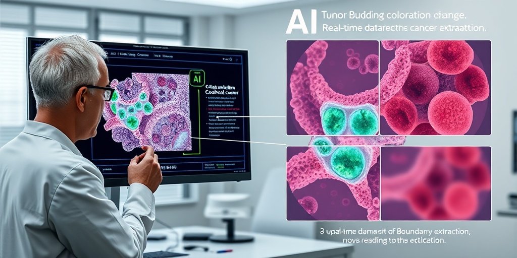
The study conducted by Peng et al. focuses on developing a full-automated TB annotation approach to assist pathologists in grading tumor budding in colorectal cancer. By extracting tumor invasive front boundaries and annotating TBs in patches, the researchers aimed to streamline the grading process and reduce the time required for assessment.
📈 Results
The automated approach achieved impressive results, with AUCs of 0.988, 0.921, and 0.929 for tumor invasive front boundary extraction across three validation datasets. Additionally, the recall rates for TB annotations were recorded at 0.850, 0.753, and 0.720. The average time taken for TB grade assessment was notably efficient, averaging around 21 seconds per WSI.
🌍 Impact and Implications
The findings from this study have significant implications for the field of pathology, particularly in the diagnosis and grading of colorectal cancer. By providing a reliable and efficient tool for TB assessment, this automated approach can enhance the accuracy of diagnoses and potentially improve treatment outcomes. The integration of such technologies in clinical practice could lead to more standardized and objective assessments in cancer pathology.
🔮 Conclusion
This study highlights the transformative potential of automation in pathology, particularly in the assessment of tumor budding in colorectal cancer. With its high accuracy and efficiency, the proposed approach represents a significant advancement in the field, offering a promising tool for pathologists. Continued research and development in this area could further enhance diagnostic capabilities and patient care in oncology.
💬 Your comments
What are your thoughts on the use of automated systems in pathology? Do you believe this technology could change the landscape of cancer diagnosis? 💬 Share your insights in the comments below or connect with us on social media:
A full-automated tumor budding annotation approach in hematoxylin and eosin-stained whole slide images of colorectal cancer.
Abstract
Accurate and efficient grade assessment of tumor budding (TB) in hematoxylin and eosin-stained whole slide images (H&E-stained WSIs) of colorectal cancer (CRC) remains challenging. This study proposes a full-automated TB annotation approach to assist in manual grade assessment by extracting tumor invasive front boundaries, annotating TBs in tumor invasive front patches, and transferring annotations to WSIs. Our approach demonstrates exceptional performance in tumor invasive front boundary extraction, achieving AUCs of 0.988, 0.921, and 0.929 on three different validation datasets. For TB annotations in tumor invasive front patches, the approach shows better recalls of 0.850, 0.753, and 0.720 on the same datasets. The average time of TB grade assisted by the approach in each WSI from different datasets is limited to 21 s, 15 s, and 18 s, respectively. These results demonstrate that this approach significantly improves assessment efficiency while guaranteeing accuracy, offering a reliable tool for CRC clinicopathological diagnosis.
Author: [‘Peng S’, ‘Chang X’, ‘Luo Y’, ‘Tang H’, ‘Yang M’, ‘Zhong C’, ‘Deng C’, ‘Hao S’, ‘Jin Y’, ‘Li M’, ‘Bai X’, ‘Nie X’]
Journal: NPJ Precis Oncol
Citation: Peng S, et al. A full-automated tumor budding annotation approach in hematoxylin and eosin-stained whole slide images of colorectal cancer. A full-automated tumor budding annotation approach in hematoxylin and eosin-stained whole slide images of colorectal cancer. 2025; (unknown volume):(unknown pages). doi: 10.1038/s41698-025-01238-4