⚡ Quick Summary
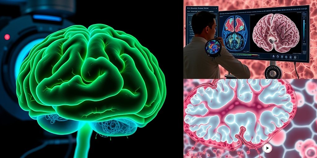
This article explores the use of Second Harmonic Generation (SHG) microscopy for brain imaging, highlighting its ability to visualize pathological processes in thick tissue samples without staining. The technique shows promise for studying conditions such as brain trauma, glioma, and Alzheimer’s disease.
🔍 Key Details
- 🔬 Technique: Second Harmonic Generation (SHG) microscopy
- 🧠 Applications: Imaging of brain trauma, fibrosis, tumorigenesis, and neuroimplant responses
- 🌟 Additional Methods: Third Harmonic Generation (THG) combined with SHG for enhanced imaging
- 📊 Focus Areas: Astrocytes, nerve fiber microtubules, and blood vessel walls
🔑 Key Takeaways
- 🔍 SHG microscopy allows for imaging of thick brain tissues without the need for staining.
- 💡 It provides insights into critical pathological processes such as brain trauma and tumorigenesis.
- 🧬 The technique is effective for visualizing collagen and noncentrosymmetric protein fibrils.
- 🤖 Recent advancements include the integration of artificial intelligence for data analysis.
- 🌍 The study reviews applications in various conditions including ischemia and neuroimplantation.
- 📈 SHG microscopy represents a breakthrough in the field of brain imaging methodologies.
📚 Background
Traditional microscopy techniques for brain imaging often struggle with issues of contrast and depth, particularly in thick tissue samples. The development of non-linear optical techniques like SHG microscopy offers a new avenue for researchers to visualize complex biological structures in their native states, paving the way for significant advancements in understanding brain pathologies.
🗒️ Study
The authors conducted a comprehensive review of the current state of SHG microscopy in brain imaging, focusing on its applications in various pathological conditions. They examined how SHG can be utilized to study brain and spinal cord injuries, gliomas, and neurodegenerative diseases, emphasizing its potential to provide detailed insights into the underlying mechanisms of these conditions.
📈 Results
The findings indicate that SHG microscopy is particularly effective in resolving the microstructure of blood vessel walls and the interactions between astrocytes and nerve fibers. The combination of SHG with THG enhances the resolution and detail of the images obtained, allowing for a more comprehensive understanding of the brain’s architecture and pathology.
🌍 Impact and Implications
The implications of this research are profound. By utilizing SHG microscopy, researchers can gain a deeper understanding of critical brain conditions, potentially leading to improved diagnostic and therapeutic strategies. The integration of artificial intelligence in analyzing SHG data further enhances the capability to interpret complex imaging results, which could revolutionize the field of neuroimaging.
🔮 Conclusion
This study highlights the transformative potential of Second Harmonic Generation microscopy in brain imaging. As researchers continue to explore its applications, we can expect significant advancements in our understanding of brain pathologies and the development of innovative treatment approaches. The future of brain imaging looks promising with these cutting-edge technologies!
💬 Your comments
What are your thoughts on the advancements in brain imaging technologies? We would love to hear your insights! 💬 Share your comments below or connect with us on social media:
Second harmonic generation for brain imaging: pathology-related studies.
Abstract
Microscopy of the brain has been facing problems of contrast and thick tissue imaging. Second harmonic generation (SHG) is a non-linear effect of the light interaction with the imaged material, resulting in photon emission at half the wavelength of the absorbed light. SHG microscopy provides an unprecedented opportunity for imaging collagen and other noncentrosymmetric protein fibrils in unstained thick tissue samples and in the live brain via a regular multiphoton setup. This opens a remarkable methodological window for imaging pathological processes of high importance, including brain trauma, fibrosis, tumorigenesis, and neuroimplant-induced foreign body response. Moreover, SHG is a valuable tool for imaging astrocytes and nerve fiber microtubules. Third harmonic generation enhanced by three-photon resonance with the Soret band of hemoglobin is combined with SHG to resolve the microstructure of blood vessel walls and astrocyte-process endfeet on gliovascular interfaces. Here, we review current state-of-the-art methods in the field of brain imaging applications of SHG, including research on brain and spinal cord injury, glioma, ischemia, Alzheimer’s disease, neuroimplantation, and brain meninges. We then address the method development perspective in the broader context of other tissue pathologies. Finally, we account for recent progress in artificial intelligence applications for SHG microscopy data analysis.
Author: [‘Paveliev M’, ‘Melnikova A’, ‘Samigullin DV’, ‘Egorchev AA’, ‘Titova AA’, ‘Kiyasov AP’, ‘Popova IY’, ‘Parpura V’, ‘Aganov AV’]
Journal: Biophys Rev
Citation: Paveliev M, et al. Second harmonic generation for brain imaging: pathology-related studies. Second harmonic generation for brain imaging: pathology-related studies. 2025; (unknown volume):(unknown pages). doi: 10.1007/s12551-025-01370-2