⚡ Quick Summary
This review highlights the role of quantitative imaging in the diagnosis and management of interstitial lung disease (ILD), emphasizing its ability to objectively measure lung abnormalities. The integration of artificial intelligence and advanced imaging techniques promises to enhance patient outcomes through personalized treatment strategies.
🔍 Key Details
- 📊 Focus: Quantitative imaging for interstitial lung disease (ILD)
- 🧩 Imaging Modalities: Primarily CT, with emerging MRI techniques
- ⚙️ Technologies: Quantitative CT, artificial intelligence, deep learning
- 🏆 Applications: Evaluation of fibrotic ILDs, including idiopathic pulmonary fibrosis and hypersensitivity pneumonitis
🔑 Key Takeaways
- 📈 Quantitative imaging allows for objective measurement of lung abnormalities.
- 💡 Early disease evaluation is enhanced through accurate classification and identification of subtle changes.
- 🤖 AI advancements are driving the development of new imaging tools.
- 🏥 Personalized treatment decisions can be made based on detailed imaging results.
- 🌍 The study emphasizes the importance of familiarity with quantitative imaging for radiologists.
- 🔬 Emerging MRI techniques may further improve ILD assessment.
- 📅 Published: 2025 in Radiol Cardiothorac Imaging.
📚 Background
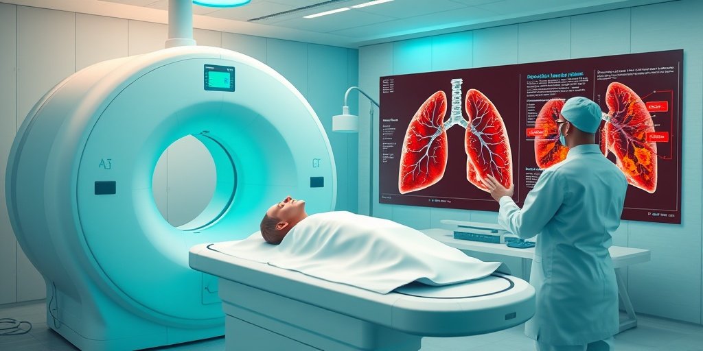
Interstitial lung disease (ILD) encompasses a diverse group of lung disorders characterized by inflammation and scarring of lung tissue. Traditional imaging methods often fall short in providing the detailed insights necessary for accurate diagnosis and treatment planning. The advent of quantitative imaging offers a promising solution, enabling clinicians to assess lung abnormalities with greater precision and reproducibility.
🗒️ Study
This review article synthesizes current knowledge on the applications of quantitative CT in evaluating fibrotic ILDs, such as idiopathic pulmonary fibrosis and hypersensitivity pneumonitis. The authors discuss the evolution of imaging technologies, including the integration of artificial intelligence and machine learning, which are enhancing the capabilities of radiologists in diagnosing and managing these complex conditions.
📈 Results
The findings underscore the effectiveness of quantitative imaging in identifying both global and regional lung abnormalities. By leveraging advanced imaging techniques, clinicians can detect subtle changes that may indicate disease progression or response to therapy, ultimately leading to improved patient outcomes.
🌍 Impact and Implications
The integration of quantitative imaging into routine clinical practice is poised to transform the landscape of ILD management. As these technologies become more prevalent, they will facilitate more accurate diagnoses and tailored treatment plans, enhancing the overall quality of care for patients with interstitial lung disease. The ongoing development of AI-driven imaging tools will further solidify the role of quantitative imaging in clinical radiology.
🔮 Conclusion
The review highlights the significant potential of quantitative imaging in the evaluation and management of interstitial lung disease. As imaging technologies continue to evolve, they promise to deliver more precise assessments, leading to better-informed treatment decisions and improved patient outcomes. The future of ILD management looks promising with the integration of these advanced imaging techniques.
💬 Your comments
What are your thoughts on the advancements in quantitative imaging for interstitial lung disease? We would love to hear your insights! 💬 Share your comments below or connect with us on social media:
Quantitative Imaging for Interstitial Lung Disease.
Abstract
Quantitative imaging has emerged as a promising tool for the diagnosis, classification, and prognostication of interstitial lung disease (ILD). Both global and regional lung abnormalities can be objectively and reproducibly measured using quantitative imaging, which is particularly useful for early disease evaluation and assessment of subtle changes. Accurate ILD classification and identification of inconspicuous changes allow for more personalized treatment decisions and, ultimately, improved patient outcomes. Because CT is the primary imaging modality for ILD evaluation, most of the computer-aided support systems have been developed for this modality and are referred to as quantitative CT. While CT continues to advance with functional capability using dual-energy technology, new MRI techniques are being developed that offer the ability to further improve ILD evaluation. Recent advancements in the field of artificial intelligence underly the development of these new quantitative imaging tools. As quantitative imaging for ILD evaluation becomes more common, it will likely play an increasingly important role in the general clinical radiology workflow, necessitating a familiarity of its use for the general radiologist. This review summarizes current applications of quantitative CT in the evaluation of fibrotic ILDs, including idiopathic pulmonary fibrosis, hypersensitivity pneumonitis, and connective tissue disease-related ILD, and highlights emerging quantitative MRI techniques for ILD assessment. Keywords: Applications-CT, Deep Learning, Machine Learning, Radiomics, CT-Quantitative, Thorax, Lung © RSNA, 2025.
Author: [‘Anderson CM’, ‘Singh R’, ‘Koo CW’]
Journal: Radiol Cardiothorac Imaging
Citation: Anderson CM, et al. Quantitative Imaging for Interstitial Lung Disease. Quantitative Imaging for Interstitial Lung Disease. 2025; 7:e250041. doi: 10.1148/ryct.250041