⚡ Quick Summary
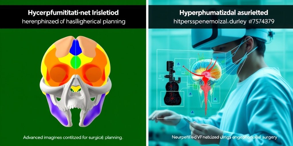
This review explores sphenoid sinus hyperpneumatization, a complex anatomical variant that can significantly impact surgical approaches to the skull base. Utilizing advanced imaging techniques and AI-driven analyses, the study highlights the potential risks and complications associated with this condition, paving the way for improved surgical outcomes.
🔍 Key Details
- 📊 Focus: Sphenoid sinus hyperpneumatization and its anatomical variants
- 🧬 Molecular Insights: Involvement of RANKL/OPG, SHH, and BMP signaling pathways
- ⚙️ Imaging Techniques: High-resolution CT (HRCT), dual-energy CT (DECT), and 7T MRI
- 🏥 Clinical Relevance: Potential complications in transsphenoidal surgery
🔑 Key Takeaways
- 🔍 Anatomical Variability: Sphenoid sinus hyperpneumatization can extend into critical areas, altering neurovascular landmarks.
- ⚠️ Surgical Risks: Increased risk of internal carotid artery injury, optic nerve impingement, and cerebrospinal fluid leaks.
- 🧠 Advanced Imaging: High-resolution imaging provides better visualization of anatomical variants, enhancing surgical planning.
- 🤖 AI Integration: Future research should incorporate AI-driven morphometric analyses for better classification and understanding.
- 🔬 Molecular Mechanisms: The study delves into the embryological basis of sinus development and its regulatory pathways.
- 🛠️ Surgical Innovations: AR/VR-assisted neuronavigation and hydroxyapatite-based techniques may reduce surgical risks.
- 📈 Clinical Implications: Understanding these variants is crucial for improving surgical precision and patient safety.
- 🌐 Research Direction: Calls for longitudinal studies to further explore the pathophysiology of hyperpneumatization.

📚 Background
The sphenoid sinus is a vital component of the skull base, characterized by its complex anatomy and significant variability among individuals. Hyperpneumatization refers to the abnormal expansion of air cells within the sphenoid sinus, which can lead to critical alterations in surrounding neurovascular structures. Despite its clinical importance, this anatomical variant is often overlooked in routine imaging, necessitating a deeper understanding of its implications for surgical practice.
🗒️ Study
This comprehensive review synthesizes primary research findings with existing anatomical and clinical knowledge to provide a thorough examination of sphenoid sinus hyperpneumatization. The authors investigate the embryological development of the sinus and the molecular mechanisms that govern its pneumatization, emphasizing the need for enhanced imaging techniques to identify these variants effectively.
📈 Results
The study demonstrates that advanced imaging modalities, such as HRCT, DECT, and 7T MRI, offer unparalleled visualization of hyperpneumatized variants. These techniques allow for the identification of thinned bony walls and high-risk zones, which are crucial for planning safe surgical interventions. The findings underscore the importance of recognizing these anatomical variations to mitigate potential surgical complications.
🌍 Impact and Implications
The insights gained from this review have significant implications for skull base surgery. By integrating advanced imaging and AI technologies, surgeons can enhance their understanding of sphenoid sinus hyperpneumatization, leading to improved surgical outcomes and reduced risks. This research not only highlights the need for a more nuanced approach to anatomical variants but also sets the stage for future innovations in surgical techniques and patient care.
🔮 Conclusion
This study emphasizes the critical role of understanding sphenoid sinus hyperpneumatization in the context of skull base surgery. By combining anatomical knowledge with cutting-edge technology, we can better navigate the complexities of this condition, ultimately enhancing surgical precision and patient safety. The future of research in this area looks promising, with the potential for AI-driven analyses to unlock further insights into its pathophysiology.
💬 Your comments
What are your thoughts on the implications of sphenoid sinus hyperpneumatization for surgical practice? We invite you to share your insights and engage in a discussion! 💬 Leave your comments below or connect with us on social media:
Sphenoid sinus hyperpneumatization: anatomical variants, molecular blueprints, and AI-augmented roadmaps for skull base surgery.
Abstract
The sphenoid sinus is a complex part of the skull base that has a high degree of anatomical variation, the most interesting of which occurs with hyperpneumatization, in which pneumatized air cells extend beyond their normal limits into the clivus, pterygoid processes, and sphenoidal wings. These hard to note hyperpneumatized imaging variants are disregarded in routine imaging but have potential to grossly alter important neurovascular landmarks, which is a challenge for the precision and safety of transsphenoidal surgical approaches. In this review, we provide an exten- sive, state-of-the-art investigation of sphenoid sinus hyperpneumatization, synthesizing novel pri- mary research discoveries with primordial radiological, anatomical, and clinical intrepidity. Our exploration to unravel the embryological basis for sinus development elicits an intricate balancing act between osteoclastic activity and the myriads of molecular actors such as RANKL/OPG, SHH, and BMP signaling pathways that delineate pneumatization in the skull base system. We demon- strate via in-depth radiological analysis how high-resolution CT (HRCT), dual-energy CT (DECT), and 7T MRI furnish unparalleled visualization of these variants, allowing identification of involved thinned bony walls, dehiscent canals, and high-risk zones for neurovascular insults. Clinically hy- perpneumatization is not just an anatomical curiosity, it may foreshadow operative complications and neurological symptoms. We discuss how it complicates endoscopic transsphenoidal ap- proaches and may increase the risk of internal carotid artery (ICA) injury, optic nerve impingement, and cerebrospinal fluid (CSF) leak. Surgical advances such as AR/VR-assisted neuronavigation and hydroxyapatite-based skull base reinforcement techniques are explored for their potential to de-risk these procedures and improve outcomes. Proactively, we propose that the future of sphenoid sinus hyperpneumatization research be one that adopts AI-driven morphometric analyses, clinically standardized classification systems, and longitudinal clinical studies to dissect its pathophysiolog- ical mysteries. This paper aims to develop an understanding of this omitted but clinically important anatomical variant by integrating basic anatomical principles with technology in order to provide clinicians, researchers, and surgical teams with a more nuanced, applicable exploration of the topic.
Author: [‘Baloiu AI’, ‘Filipoiu F’, ‘Toader C’, ‘Covache-Busuioc RA’, ‘Munteanu O’, ‘Serban M’]
Journal: Front Endocrinol (Lausanne)
Citation: Baloiu AI, et al. Sphenoid sinus hyperpneumatization: anatomical variants, molecular blueprints, and AI-augmented roadmaps for skull base surgery. Sphenoid sinus hyperpneumatization: anatomical variants, molecular blueprints, and AI-augmented roadmaps for skull base surgery. 2025; 16:1634206. doi: 10.3389/fendo.2025.1634206