⚡ Quick Summary
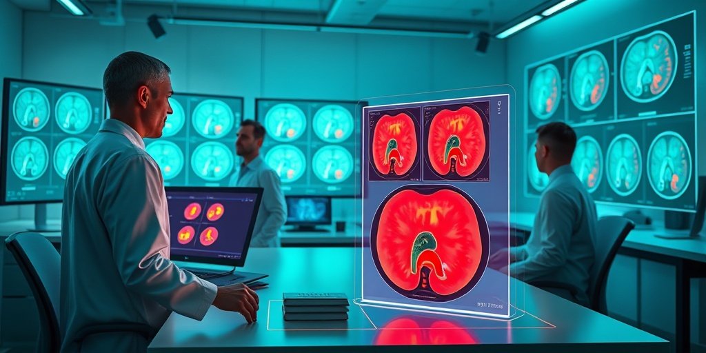
This study presents a collaborative approach between a radiologist and data scientists to enhance the detection and segmentation of rectal tumors in MR imaging. The results indicate a high level of agreement in tumor segmentation, achieving a mean Dice Similarity Coefficient (DSC) of 0.965 and a mean Jaccard Index (JI) of 0.943, paving the way for future AI-driven solutions.
🔍 Key Details
- 📊 Dataset: 37 patients with rectal cancer
- 🧩 Imaging Techniques: T2- and diffusion-weighted MR images
- 🤝 Collaboration: Radiologist and data scientists
- ⚙️ Evaluation Metrics: Dice Similarity Coefficient (DSC) and Jaccard Index (JI)
🔑 Key Takeaways
- 🤖 AI integration in medical imaging can significantly enhance tumor detection and segmentation.
- 💡 Collaboration between radiologists and data scientists is crucial for developing effective AI models.
- 📈 High accuracy achieved with a mean DSC of 0.965 and JI of 0.943 demonstrates the potential of this approach.
- 📝 Radiologist annotations play a vital role in training AI systems for accurate tumor segmentation.
- 🌟 Future implications include improved clinical workflows and patient outcomes in rectal cancer management.
- 🔍 Study conducted at Stanford University, showcasing a model for interdisciplinary collaboration.
📚 Background
Magnetic Resonance (MR) imaging is the gold standard for staging rectal cancer due to its superior soft tissue contrast. However, the segmentation of rectal tumors remains a challenge due to overlapping appearances with normal tissues and variability in imaging parameters. Traditional methods rely on multiple radiologist reviews, which can be costly and time-consuming. The integration of Artificial Intelligence (AI) offers a promising avenue for improving segmentation accuracy and efficiency.
🗒️ Study
This study aimed to create a workflow for tumor detection and segmentation by fostering collaboration between a radiologist and two data scientists. The team utilized radiologist-led training and clinical notes from Epic Electronic Health Records (EHR) to guide the data scientists in accurately identifying and segmenting rectal tumors on MR images. The segmentations were then reviewed and refined by the radiologist to ensure accuracy.
📈 Results
The collaboration yielded impressive results, with the data scientists successfully identifying rectal tumors across all evaluated MR images. The segmentations demonstrated a strong agreement with the radiologist’s edits, achieving a mean DSC of 0.965 and a mean JI of 0.943. Discrepancies were primarily due to over- or under-segmentation, highlighting the complexity of accurately delineating tumors from surrounding structures.
🌍 Impact and Implications
The findings of this study have significant implications for the future of rectal cancer management. By establishing a reliable method for generating high-quality labeled MR datasets, this collaborative approach lays the groundwork for training robust AI models. Such advancements could streamline clinical workflows, reduce costs, and ultimately improve patient outcomes in rectal cancer diagnosis and treatment.
🔮 Conclusion
This study illustrates the potential of interdisciplinary collaboration in enhancing the accuracy of tumor detection and segmentation in rectal cancer. By combining the expertise of radiologists and data scientists, we can develop AI-driven solutions that transform clinical practices. The future of rectal cancer imaging looks promising, and further research in this area is encouraged to unlock the full potential of AI in healthcare.
💬 Your comments
What are your thoughts on the integration of AI in medical imaging? We would love to hear your insights! 💬 Leave your comments below or connect with us on social media:
AI-ready rectal cancer MR imaging: a workflow for tumor detection and segmentation.
Abstract
BACKGROUND: Magnetic Resonance (MR) imaging is the preferred modality for staging in rectal cancer; however, despite its exceptional soft tissue contrast, segmenting rectal tumors on MR images remains challenging due to the overlapping appearance of tumor and normal tissues, variability in imaging parameters, and the inherent subjectivity of reader interpretation. For studies requiring accurate segmentation, reviews by multiple independent radiologists remain the gold standard, albeit at a substantial cost. The emergence of Artificial Intelligence (AI) offers promising solutions to semi- or fully-automatic segmentation, but the lack of publicly available, high-quality MR imaging datasets for rectal cancer remains a significant barrier to developing robust AI models.
OBJECTIVE: This study aimed to foster collaboration between a radiologist and two data scientists in the detection and segmentation of rectal tumors on T2- and diffusion-weighted MR images. By combining the radiologist’s clinical expertise with the data scientists’ imaging analysis skills, we sought to establish a foundation for future AI-driven approaches that streamline rectal tumor detection and segmentation, and optimize workflow efficiency.
METHODS: A total of 37 patients with rectal cancer were included in this study. Through radiologist-led training, attendance at Stanford’s weekly Colorectal Cancer Multidisciplinary Tumor Board (CRC MDTB), and the use of radiologist annotations and clinical notes in Epic Electronic Health Records (EHR), data scientists learned how to detect and manually segment tumors on T2- and diffusion-weighted pre-treatment MR images. These segmentations were then reviewed and edited by a radiologist. The accuracy of the segmentations was evaluated using the Dice Similarity Coefficient (DSC) and Jaccard Index (JI), quantifying the overlap between the segmentations delineated by the data scientists and those edited by the radiologist.
RESULTS: With the help of radiologist annotations and radiology notes in Epic EHR, the data scientists successfully identified rectal tumors in Slicer v5.7.0 across all evaluated T2- and diffusion-weighted MR images. Through radiologist-led training and participation at Stanford’s weekly CRC MDTB, the data scientists’ rectal tumor segmentations exhibited strong agreement with the radiologist’s edits, achieving a mean DSC [95% CI] of 0.965 [0.939-0.992] and a mean JI [95% CI] of 0.943 [0.900, 0.985]. Discrepancies in segmentations were attributed to over- or under-segmentation, often incorporating surrounding structures such as the rectal wall and lumen.
CONCLUSION: This study demonstrates the feasibility of generating high-quality labeled MR datasets through collaboration between a radiologist and two data scientists, which is essential for training AI models to automate tumor detection and segmentation in rectal cancer. By integrating expertise from radiology and data science, this approach has the potential to enhance AI model performance and transform clinical workflows in the future.
Author: [‘Selby HM’, ‘Son YA’, ‘Sheth VR’, ‘Wagner TH’, ‘Pollom EL’, ‘Morris AM’]
Journal: BMC Med Imaging
Citation: Selby HM, et al. AI-ready rectal cancer MR imaging: a workflow for tumor detection and segmentation. AI-ready rectal cancer MR imaging: a workflow for tumor detection and segmentation. 2025; 25:88. doi: 10.1186/s12880-025-01614-3
2 Comments
Weekly Health & AI Digest – March 20, 2025 - Yesil Science
[…] AI-ready rectal cancer MR imaging: a workflow for tumor detection and segmentation. […]
Weekly Health & AI Digest – March 20, 2025 - Yesil Science
[…] 🔹 AI-ready rectal cancer MR imaging: a workflow for tumor detection and segmentation. […]